The Antebrachium

Chapter 39The Antebrachium


Anatomy


The antebrachium lies between the elbow and carpus and is composed principally of the radius and small vestigial portion of the ulna and the flexor and extensor muscles. The tendons of the superficial and deep digital flexor muscles, the accessory ligament of the superficial digital flexor tendon, and the carpal sheath are discussed elsewhere (see Chapters 69, 70, and 75). The medial aspect of the antebrachium is relatively devoid of soft tissue coverage, and this is important when considering fractures of the radius. Major neurovascular structures include the median artery (continuation of the brachial artery in the proximal antebrachium), vein, and nerve; the radial, ulnar, and cutaneous antebrachial nerves; and the accessory cephalic and cephalic veins.


Clinical Diagnosis and Imaging Considerations


Lameness associated with the antebrachial region is relatively unusual. Clinical signs are obvious in horses with unstable radial fractures or in those with marked soft tissue swelling. In others diagnosis can be challenging, and ruling out other causes of forelimb lameness and then using diagnostic imaging to reach a definite diagnosis may be necessary. Perineural analgesia of the median and ulnar nerves (see Chapter 10) is performed to rule out more distal sources of pain.


Definitive diagnosis of most lameness problems of the antebrachium can be made using conventional radiography and ultrasonography, but nuclear scintigraphy is useful for diagnosing incomplete and stress fractures of the radius, enostosis-like lesions, and enthesopathy at the origin of the accessory ligament of the superficial digital flexor tendon.


Osteochondroma of the Distal Aspect of the Radius


See Chapter 75 for a discussion of osteochondroma of the distal aspect of the radius.


Traumatic Physitis and Closure of the Distal Radial Physis


A syndrome of vague forelimb lameness believed to be associated with inflammation or pain originating from the distal radial physis has been recognized in young racehorses in early training. Anecdotally the condition appears to be more prevalent in 2-year-old colts. Distal radial physeal closure determined radiologically occurred earlier in fillies (701 days) than in colts (748 days).1 Presumably the condition results from repetitive trauma to an open physis. The term open knees is commonly used to describe the state of skeletal immaturity. This condition is distinct from physeal dysplasia (physitis), because the condition is not a developmental abnormality, is not associated with clinically apparent enlargement of the metaphyseal region, and occurs in 2-year-old horses in active training.


Mild to moderate forelimb lameness is vague, without indications of a problem elsewhere in the limb. The condition is usually bilateral, but horses can show unilateral lameness, because one limb is more painful than the other. Horses often have a choppy stride, with the legs carried wide. Focal heat may be present, but swelling is usually absent or minimal, and pain may be difficult to detect. Presumptive diagnosis is made on the basis of history, clinical signs, and ruling out other causes of lameness. A positive response to median and ulnar nerve blocks can be used to confirm that the distal aspect of the antebrachium is the source of pain, but this can also abolish subchondral bone pain in the carpus. Definitive radiological abnormalities are rarely present, but a radiologically open physis supports the diagnosis. Nuclear scintigraphy is generally not helpful, because all horses of this age have moderate-to-intense increased radiopharmaceutical uptake (IRU) at the physis. However, scintigraphy is useful for identifying or ruling out other potential causes of lameness, and asymmetrical radiopharmaceutical uptake (greater in the affected physis of the more severely affected limb) may support the diagnosis.


Treatment consists primarily of rest or a reduction in exercise intensity and systemic nonsteroidal antiinflammatory drugs (NSAIDs). Duration of rest varies with the skeletal maturity of the horse and severity of the condition. Local injection of corticosteroids or other drugs, such as homeopathic remedies, and systemic treatment with anabolic steroids have been used but are of dubious value. For some horses, slow jogging for 4 to 6 weeks may be all that is required; for others, stall confinement with hand-walking exercise progressing to paddock turnout for several months may be necessary. Follow-up radiographs can be used to monitor physeal closure, which is often used to determine the appropriate time to resume harder training.


The issue of distal radial physeal closure, the role of radiographs in making this determination, and how to determine the point when training should commence are controversial. Many trainers and veterinarians customarily obtain radiographs of the distal aspect of the radius of 2-year-olds, and those with open knees (radiological evidence that bony union at the physis is incomplete) are withheld from hard training until the physes have closed. Radiological closure of the distal radial physis generally occurs by 20 to 24 months of age2 or slightly later.1 Scintigraphic activity of the distal radial physis persists well after radiological evidence of closure is observed.3 However, endochondral ossification ceases (biological closure) before fusion is evident radiologically, and in our experience horses with a thin or faintly visible physeal remnant visible radiologically are at low risk for traumatic physitis.


Because the diagnosis is difficult to substantiate and pain may originate from an undetermined source, giving an accurate prognosis is difficult. If other more common conditions have been ruled out and the diagnosis of traumatic physitis is accurate, the prognosis is excellent. Many horses remaining in training develop signs of carpal lameness, and traumatic physitis may simply represent a prodromal phase of early osteoarthritis and bone pain. Finally, no correlation between age or month of closure of the distal radial physes and money won, races won, fastest mile, or fastest win mile during the 2-year-old year was found in Standardbreds.4


Radial Fractures


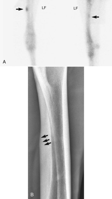
Radial fractures almost always result from external trauma, often a kick from another horse in adults, or from being stepped on or kicked by a mare in foals. Stress fractures of the radius also occur,5,6 but in our experience true stress fractures of the radius are rare, and the description of those reported by others is similar to what we have termed enostosis-like lesions (see the following discussion). One of us (MWR) recently evaluated images of a 4-year-old Thoroughbred colt in race training, with a history of sudden left forelimb lameness, with a genuine radial stress fracture involving the medial, middiaphyseal cortex (Figure 39-1). Cortical location of stress fractures as determined scintigraphically and radiologically differentiates this injury from enostosis-like lesions.


Clinical signs depend on the severity and location of the fracture. Horses with complete fractures (which are nearly always displaced) are severely lame (non–weight bearing, grade 5) and have marked soft tissue swelling associated with the fracture itself or the site of the original wound. The limb may have an unusual angle, and crepitus is usually audible and palpable. Often an associated wound results from the initial injury or is caused by fragment penetration, especially on the medial aspect of the antebrachium. Horses with incomplete or nondisplaced fractures have moderate to severe lameness (grade 3 to 5) shortly after the injury, but within 12 to 72 hours they are often fully weight bearing and walking with minimal lameness. However, resumption of exercise or turnout often results in the fracture becoming displaced within 1 to 2 days (see Figure 3-4). Horses with true stress fractures have moderate lameness (grade 1 to 3) at a trot.


For horses with complete, displaced (unstable) fractures of the radius the diagnosis is straightforward. Radiology is needed only to define fracture configuration and to determine if repair is possible. Radiographs are essential in the initial evaluation of any horse with a wound in the antebrachium or over the proximal aspect of the carpus that has a history of acute, moderate-to-severe lameness associated with the injury. Lameness associated with incomplete or hairline fractures of the radius may be transient, but radiographs often reveal obvious or suspicious fracture lines (Figure 39-2, A). Any radiological evidence of bone injury, often a localized cortical fragmentation or compression fracture, warrants high suspicion of an incomplete fracture, and a full series of radiographs should be obtained. Any horse that has persistent lameness after antebrachial trauma in which original radiological findings were negative should be reevaluated within 7 to 10 days, when a fracture may be evident. The horse should be confined to box rest in the interim. Diagnosis in horses with incomplete fractures or stress fractures can sometimes be difficult. Scintigraphic examination is important to differentiate fracture from other problems of the radius, such as enostosis-like lesions.

Only gold members can continue reading. Log In or Register to continue

Stay updated, free articles. Join our Telegram channel

Jun 4, 2016 | Posted by in EQUINE MEDICINE | Comments Off on The Antebrachium

Full access? Get Clinical Tree

Get Clinical Tree app for offline access