The acute abdomen

29 The acute abdomen


Differential diagnoses of acute abdomen are discussed in Chapter 8 and some of the most common disorders are discussed in greater detail here using case examples.


Septic (Bacterial) Peritonitis


Theory refresher


Free peritoneal fluid and gas


Abdominal palpation is an insensitive means of detecting free peritoneal fluid. Ultrasonography is extremely useful in this respect even in relatively inexperienced hands. Small amounts of free peritoneal fluid are most readily detected at the apex of the urinary bladder and between liver lobes. If ultrasonography is not available, loss of serosal detail may be identified radiographically.


Abdominal radiography is useful for identifying free peritoneal gas (often most readily seen between the liver and diaphragm) as well as diagnosing intestinal foreign body material and obstruction. Radiography will be less useful in this respect if a large volume of free peritoneal fluid is present due to increased loss of contrast.


In animals without a history of recent (last 3–4 weeks) laparotomy or open system abdominocentesis, free peritoneal gas suggests rupture of a hollow viscus and is an indication for emergency surgical exploration. This also applies to animals that have suffered penetrating injuries as free peritoneal gas implies full thickness penetration into the abdomen.


Case example 1 – surgical gastrointestinal wound dehiscence


Case management


Morphine (0.3 mg/kg slow i.v.) was administered for analgesia. An abdominal ultrasound was performed using a focused assessment with sonography for trauma (FAST) protocol. This involved scanning the dog in lateral recumbency and obtaining both transverse and longitudinal views at each of four sites – just caudal to the xiphoid process (last sternebra), on the midline over the urinary bladder, and at the right and left flank regions. Despite this thorough evaluation, no peritoneal fluid could be identified. However, given the dog’s history and clinical signs, there was a very high index of suspicion for bacterial peritonitis secondary to surgical gastrointestinal wound dehiscence and a diagnostic peritoneal lavage (DPL) was therefore performed (see p. 294).


The peritoneal fluid subsequently obtained was centrifuged and smears made from the sediment. Cytology demonstrated degenerate neutrophils with occasional intracellular rod-shaped bacteria and a diagnosis of bacterial peritonitis was therefore confirmed. A peritoneal fluid glucose concentration of less than 2.8 mmol/l is reported to have high specificity for bacterial peritonitis and was found to be 2.0 mmol/l in this case.


Intravenous antibiosis using amoxicillin/clavulanic acid (20 mg/kg) was commenced and an exploratory laparotomy performed. This revealed dehiscence with leakage from the enterotomy incision that was debrided and repaired. Antibiosis was continued postoperatively, first intravenously and then per os, and culture of the fluid obtained from the abdomen following DPL revealed a scant growth of Escherichia coli that was sensitive to amoxicillin/clavulanic acid. The dog made an uneventful recovery from the surgery and was discharged after 5 days to complete a 2-week course of antibiotics.


Case example 2 – uterine rupture


Major body system examination


On presentation the dog was obtunded. Cardiovascular examination revealed a heart rate of 200 beats per minute with very weak femoral pulses and absent dorsal pedal pulses. Mucous membranes were hyperaemic with a rapid capillary refill time. Respiratory examination revealed tachypnoea with appropriate lung auscultation. The abdomen was tense and painful on palpation but a more thorough examination could not be performed due to the dog’s recumbency. Marked pyrexia (rectal temperature 40.4°C) was identified. The vulva was swollen but no discharge was detected. A brown-tinged serous mammary discharge was present but the mammary glands were not enlarged or obviously inflamed.


Haemoabdomen


Causes of haemoabdomen are listed in Box 29.1.


Theory refresher


Canine haemangiosarcoma


Canine haemangiosarcoma is most commonly diagnosed in German shepherd dogs followed by golden retrievers. Other large breed dogs, such as Labrador retrievers and Boxers, are also over represented. The spleen is the most common site of origin but right atrial, pericardial and hepatic canine haemangiosarcoma are also reported relatively frequently. Any site in the body that contains vascular endothelium can theoretically be affected.


Haemangiosarcoma is presumed to have metastasized at the time of diagnosis in the vast majority of cases, regardless of whether or not metastases are detected by currently available imaging modalities. The prognosis is very poor, with survival times in the region of 2–4 months reported for splenic haemangiosarcoma following splenectomy alone; survival may in some cases be improved modestly by additional chemotherapy and immunotherapy.


In dogs presenting with haemoabdomen presumed to be secondary to rupture of a splenic lesion, splenectomy will be palliative and remove the risk of recurrent haemorrhage in the short term. It should be remembered that the nature (benign or malignant) of hepatic nodules that may be identified in such cases cannot be determined based on ultrasonographic appearance and surgical intervention therefore remains a rational choice. Palliative surgery is unlikely to be an option in primary hepatic haemangiosarcoma.


Abdominal counterpressure bandage


The purpose of an abdominal bandage is to apply external pressure that will increase intra-abdominal pressure and thereby tamponade bleeding. Realistically, intra-abdominal pressure is only likely to be increased sufficiently to exceed venous pressure and reduce venous haemorrhage. Therefore, excessively tight bandages do not offer any significant advantage and are likely to be associated with greater complications.


Traditionally a soft material has been used as the primary layer of an abdominal pressure bandage with an elastic material applied on top. In the author’s experience, however, these bandages are more likely to slip and the author prefers to use an elastic adhesive primary layer despite the inherent difficulties in subsequent removal. The bandage should be placed starting caudally, approximately at the level of the pubis, and moving in a cranial direction up to the xiphoid, stopping before the caudal rib margin.


The bandage should be removed once the patient has remained stable for a reasonable period. Removal should commence at the cranial end (i.e. in the opposite direction to how the bandage was placed) and should be staggered by cutting a small section every 30–60 minutes over a period of several hours. The patient should be monitored closely for signs of deterioration during removal of counterpressure.


Abdominal bandages should not be used in animals with respiratory compromise or those suffering from diaphragmatic rupture.


Case example 3


Case management


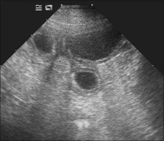
Aggressive intravenous fluid resuscitation with an isotonic crystalloid solution was commenced (60 ml/kg bolus via pressure infusor). Abdominal ultrasonography revealed the presence of a large volume of echogenic peritoneal fluid. 
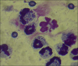
Abdominocentesis (see p. 293) was performed and the non-clotting haemorrhagic fluid was found to have a PCV of 30%; cytology of the fluid revealed red blood cells, occasional erythrophagocytosis, no platelets and no evidence of sepsis. An abdominal counterpressure bandage was applied and fluid therapy discontinued as soon as the dog was assessed as being adequately perfused. The dog remained stable subsequently and the abdominal bandage was removed gradually over several hours.


The following day the dog was referred for further management. Abdominal ultrasonography revealed a cavitary splenic lesion and although the liver was hyperechoic, no focal lesions were identified. Thoracic radiography was performed under general anaesthesia and found to be unremarkable. Complete splenectomy and a liver biopsy were then carried out, from which the dog made an uneventful recovery. Histopathology confirmed the suspicion of splenic haemangiosarcoma although no neoplastic cells were identified in the sample of liver submitted.


Uroabdomen


Sep 3, 2016 | Posted by in SMALL ANIMAL | Comments Off on The acute abdomen

Full access? Get Clinical Tree

Get Clinical Tree app for offline access