19 Pallor and approach to anaemia
Pallor
In many cases it will be possible on the basis of historical and physical examination findings to determine whether anaemia or hypovolaemia may be present. Concomitant hypovolaemia and anaemia are typically seen where anaemia is secondary to acute blood loss and the degree of pallor may be more severe than would otherwise be expected for the degree of hypovolaemia. It is important to remember that the packed cell volume may be normal for several hours following acute blood loss in dogs (but serum total solids should be low) (see Ch. 3).
Anaemia
A rational approach to the investigation of anaemia starts with classification of the anaemia as regenerative (due to red blood cell loss or destruction) or nonregenerative (due to reduced red blood cell production). In the emergency setting, this is done on the basis of peripheral blood smear examination, with regenerative anaemia being manifested as polychromasia, anisocytosis and a possible increase in nucleated red blood cells (see Figures 3.3 and 3.5). Regenerative anaemia occurs as a result of haemolysis or haemorrhage, and additional findings are used to determine which of these two processes is the likely cause (Figure 19.1).
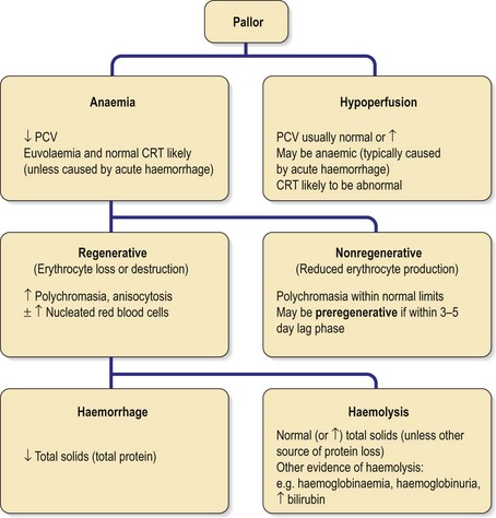
Figure 19.1 Algorithm for pallor and approach to anaemia. CRT, capillary refill time; PCV, packed cell volume.
Clinical Tip
Regenerative anaemia
Causes of regenerative anaemia are listed in Box 19.1.


