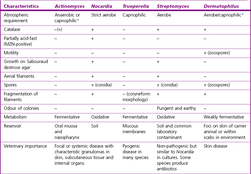
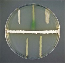
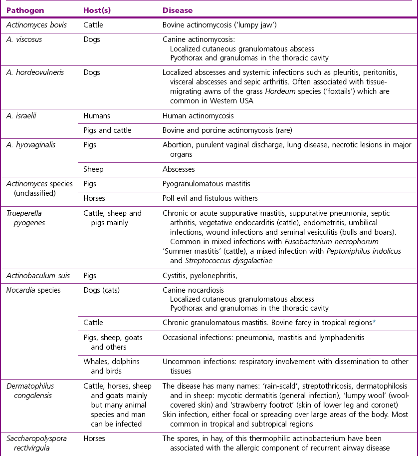
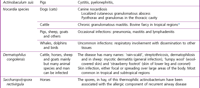
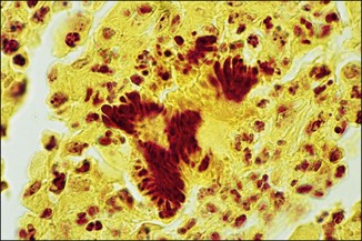
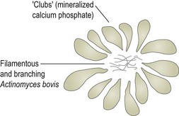
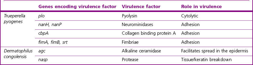
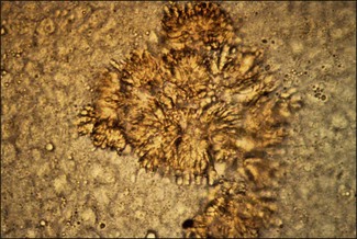
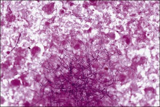
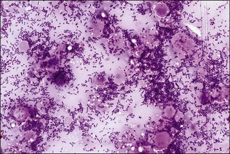
Chapter 10 The class Actinobacteria comprises a heterologous group of procaryotes, containing some genera which have the ability to form Gram-positive, branching filaments of less than 1 μm in diameter. Thus, these bacteria were originally thought to be fungi. However, fungi are eucaryotes and their filaments (hyphae) are always greater than 1 μm in width. The main animal pathogens in the Actinobacteria are found in the genera Actinomyces, Actinobaculum and Trueperella in the family Actinomycetaceae, the genera Mycobacterium, Corynebacterium, Rhodococcus and Nocardia in the family Corynebacteriaceae and in the genus Dermatophilus. Members of the genus Nocardia are closely related to Corynebacterium, Mycobacterium and Rhodococcus species but Actinomyces differs from these in its DNA guanine/cytosine ratio and in the chemical composition of its cell wall. Information about the genera Corynebacterium and Rhodococcus is presented in Chapter 9 and the mycobacteria are discussed in Chapter 11. Other genera included in the Actinobacteria are Streptomyces and Actinomadura; some species of each produce mycetomas in humans but are rarely pathogenic for animals. Streptomyces species are prolific producers of antimicrobial substances (Fig. 10.1) and are common contaminants on laboratory agar media. Species in the genera Saccharopolyspora and Thermoactinomyces are not invasive but inhalation of their spores can cause allergic pulmonary disease in man and horses and possibly in other domestic animals fed or exposed to mouldy hay. The general characteristics of the genera Actinomyces, Nocardia, Trueperella, Streptomyces and Dermatophilus are presented in Table 10.1. Table 10.1 General features of Actinomyces, Nocardia, Trueperella, Streptomyces (non-pathogenic) and Dermatophilus species *capnophilic (carboxyphilic) = carbon dioxide required for maximum growth, as opposed to microaerophilic, used to describe an organism that requires a reduced oxygen tension,. + = positive reaction, − = negative, −(+) = most species are negative (a few positive) Figure 10.1 Streptomyces species (horizontal streak) on Isosensitest agar demonstrating antimicrobial activity against other bacteria that were streaked to within 2 mm of the Streptomyces species: S. aureus (top left), Pseudomonas aeruginosa (top centre), Bacillus cereus (top right), Salmonella species (bottom left) and E. coli (bottom right). Pseudomonas aeruginosa is the only bacterium not inhibited by the antimicrobial factor(s) from the Streptomyces species. The pathogenic Actinobacteria and the diseases that they cause are summarized in Table 10.2. Table 10.2 Diseases caused by selected pathogenic actinobacteria of veterinary importance *some mycobacteria have also been implicated in bovine farcy Actinomyces bovis gains access to the alveolar region of the jaw in cattle from the oral cavity, probably through trauma to the mucosa. It initiates a rarefying osteomyelitis and soft tissue reaction, the condition being referred to as ‘lumpy jaw’. Bacterial colonies form in the tissues with ‘clubs’ of mineralized calcium phosphate forming around them to create microscopic ‘club colonies’ or ‘rosettes’. Figure 10.2 shows a diagram of a ‘club colony’. The club formation is the result of phosphatase activity and is a host reaction to a chronic infection. Granulation, mononuclear infiltration and fibrosis occur in the lesions with sinus tracts leading to the outside. Exudate from the tracts contains pus with ‘sulphur granules’ that are about 1–2 mm in diameter, within which club colonies can be found if the granules are crushed and examined microscopically. Strains of Actinomyces similar to A. bovis have been isolated from sows with granulomatous lesions in the mammary glands. These porcine strains show minor biochemical and antigenic differences from the bovine strains. Actinomyces hyovaginalis is associated with abortions and purulent vaginal discharge in sows and with infections in various sites in pigs and in sheep (Collins et al. 1993, Storms et al. 2002, Foster et al. 2012). A. viscosus causes a clinical syndrome in dogs indistinguishable from that initiated by Nocardia species. Two syndromes can occur, either separately or together. One is a localized granulomatous lesion involving skin and subcutis, the other is a pyothorax with granulomas in thoracic tissue and often a large accumulation of sanguineopurulent pleural fluid containing soft white granules about 1 mm in diameter. Trueperella pyogenes produces a range of virulence factors, the most significant of which is a haemolytic exotoxin, pyolysin (Table 10.3). This toxin is cytolytic for neutrophils and macrophages. Adhesion to host tissues is facilitated by neuraminidases and extracellular matrix-binding proteins. The organism also possesses fimbriae, produces proteases and can form biofilms but the exact role of these factors in disease production has not been definitively established. Nocardiae are difficult to definitively identify using phenotypic methods and many species have been reclassified in recent years based on molecular techniques. These aerobic, and essentially saprophytic, bacteria cause suppurative and pyogranulomatous reactions in immunosuppressed hosts or animals that have been exposed to large doses of the bacterium. The pathogenic nocardiae survive within phagocytic vacuoles by preventing phagolysosome formation. This is probably due to the surface lipids; Nocardia species have a cell wall similar to the mycobacteria. Genes encoding putative virulence factors have been identified in some Nocardia species but the molecular pathogenesis of nocardial infections in animals is currently unclear. Superoxide dismutases and catalases appear to be important in resistance to phagocytosis and cell lipids may provoke the characteristic granulomatous reaction. Exudates are sanguineopurulent and can sometimes contain soft granules consisting of bacteria, neutrophils and debris. They lack the microstructure of the sulphur granules produced by some of the Actinomyces species. Nocardia asteroides was previously considered to account for the majority of infections in animals but following their reclassification, a number of species are now known to be associated with animal disease (Table 10.2). Nocardiform placentitis associated with a number of different members of the Actinobacteria, in particular Amycolatopsis species and Crossiella equi, have been documented in the USA and sporadically elsewhere (Erol et al. 2012). Dermatophilus congolensis causes skin infections that can affect many animal species and humans, but the condition is most commonly seen in cattle, sheep, goats and horses and polar bears in zoological collections. The infection is characterized by the formation of thick crusts which come away easily with a tuft of hair, leaving a moist, depressed area with bleeding points from capillaries. Virulence factors include an alkaline ceramidase and a number of proteases which facilitate invasion of the epidermis (García-Sánchez et al. 2004, Norris et al. 2008). Infections can be localized but have a tendency to spread over large areas of the body and the morbidity and mortality can be high, especially in tropical regions. The position of the lesions varies with the predisposing conditions. In periods of high rainfall the lesions tend to occur along the backs of animals. Where there is a heavy infestation with Amblyomma ticks the lesions are present in the predilection sites of the ticks: dewlap, axillae, udder, scrotum and escutcheon. In the dry season, in tropical regions, when feed is scarce the lesions are on the muzzle, head and lower limbs due to the animals foraging in thorn-covered scrub. The pathogenic actinobacteria and the diseases that they cause are summarized in Table 10.2 and virulence factors of T. pyogenes and D. congolensis are given in Table 10.3. Sulphur granules are the best specimens for direct examination in infections caused by A. bovis or A. viscosus. The pus or exudate is placed in a Petri dish and washed carefully with a little distilled water to expose the yellowish sulphur granules of A. bovis (Fig. 10.3) or the softer greyish-white granules of A. viscosus. A granule is placed on a microscope slide in a drop of 10% KOH and gently crushed by applying pressure on the coverslip. The characteristic clubs can be seen if the preparation is examined under the low-power objective of a microscope. A view of a club colony, in situ, can be seen in stained histological sections (Fig. 10.4). If smears are made from the granules and stained with the Gram stain, delicate, Gram-positive, branching filaments (Fig. 10.5) can be observed. Occasionally short filaments or pleomorphic diphtheroidal forms may predominate. Figure 10.4 Club colonies in a tissue section from an A. bovis infection in a cow with lumpy jaw. (Plaut stain, ×400) Gram-stained smears of pus or mastitic milk samples in T. pyogenes infections usually reveal large numbers of small, highly pleomorphic, Gram-positive forms (Fig. 10.6). They tend to be a mixture of cocci, rods and pear-shaped cells. Occasionally short branching forms may be seen.
The Actinobacteria
Changes in Nomenclature
Previous name
Present name
Arcanobacterium (Actinomyces) pyogenes
Trueperella pyogenes
Actinomyces suis
Actinobaculum suis
Nocardia caviae
Nocardia otitidiscaviarum
Pathogenicity
Actinomyces, Trueperella and Actinobaculum species
Nocardia species
Dermatophilus congolensis
Laboratory Diagnosis of Actinomyces, Trueperella and Actinobaculum Species
Direct microscopy
< div class='tao-gold-member'>
![]()
Stay updated, free articles. Join our Telegram channel

Full access? Get Clinical Tree


The Actinobacteria
Only gold members can continue reading. Log In or Register to continue
