Chapter 81Tendon Lacerations
Diagnosis
Gross Appearance of the Wound
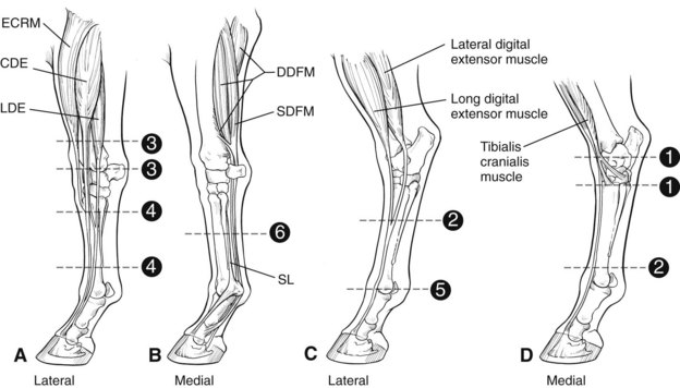
Any laceration over the dorsal or palmar/plantar surface of the limb distal to the stifle or elbow, especially across the dorsal aspect of the tarsus, distal dorsal aspect of the tarsus, dorsal metatarsal region, distal aspect of the radius, dorsal metacarpal region, and dorsal fetlock region may involve a tendon (Figure 81-1). Extensor tendons and the superficial digital flexor tendon (SDFT) are positioned directly under the skin; therefore minor-appearing wounds can transect these tendons completely. Direct visual inspection may reveal transected tendon fibers protruding from the wound. However, injuries sustained at the gallop may result in a skin wound removed from the site of tendon damage because of the movement of the skin during exercise. The position of the wound relative to synovial structures should be evaluated with care because concurrent synovial contamination or infection reduces the prognosis and necessitates specific emergency treatment.
Evaluation of Gait
Extensor Tendons
Proximal or dorsal to the tarsus, transection of the long digital extensor, cranialis tibialis, and fibularis (peroneus) tertius tendons is most frequent. ![]() If the fibularis tertius is disrupted, the hock can be extended while the stifle is flexed, indicating loss of the reciprocal apparatus. The gastrocnemius tendon develops a characteristic wrinkle in this extended position (see Figure 80-2 and page 802). The degree of gait abnormality may be mild. Transection of all the extensor tendons over the tarsus still allows full weight bearing with the foot flat on the ground. A greater tarsal extension during the swing phase of the stride and intermittent knuckling of the digit can be detected.
If the fibularis tertius is disrupted, the hock can be extended while the stifle is flexed, indicating loss of the reciprocal apparatus. The gastrocnemius tendon develops a characteristic wrinkle in this extended position (see Figure 80-2 and page 802). The degree of gait abnormality may be mild. Transection of all the extensor tendons over the tarsus still allows full weight bearing with the foot flat on the ground. A greater tarsal extension during the swing phase of the stride and intermittent knuckling of the digit can be detected.
Digital Palpation of the Wound
Digital palpation is a simple and direct way to determine the extent of damage to structures below the skin. Integrity of the tendons is readily determined by feel. Partial tears can be distinguished from complete tears, and this affects treatment (see Partial Tendon Lacerations
Stay updated, free articles. Join our Telegram channel

Full access? Get Clinical Tree


