CHAPTER 4 Temporomandibular Joint
Limited imaging of the temporomandibular joint (TMJ) may be done using a dental radiograph machine and dental film or digital sensor. Skull radiographs and computed tomography images provide more information and may be necessary in many patients to adequately evaluate the TMJs. The radiographs in this section were all done using an extraoral technique with a digital sensor and dental radiograph machine.


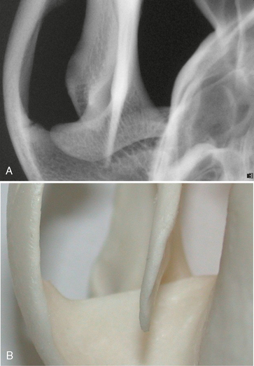
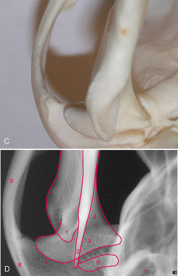
FIGURE 4-1 Left temporomandibular joint (TMJ) in a medium-size dog. A, Radiograph of the left TMJ from a dog skull, dorsovental view. B, Dorsal view of left TMJ from a prepared dog skull. C, Ventral view (mirror) of the TMJ from a prepared dog skull. D, Same radiograph as A.


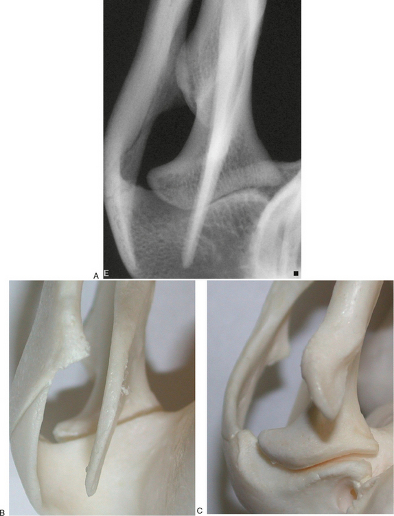
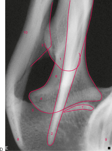
FIGURE 4-2 Left temporomandibular joint (TMJ) in a medium-size dog. A, Radiograph of the left TMJ from a dog skull, intraorbital view. B, Dorsal view of left TMJ from a prepared dog skull. C, Ventral view (mirror) of the TMJ from a dog skull. D, Same radiograph as A.
Stay updated, free articles. Join our Telegram channel

Full access? Get Clinical Tree


