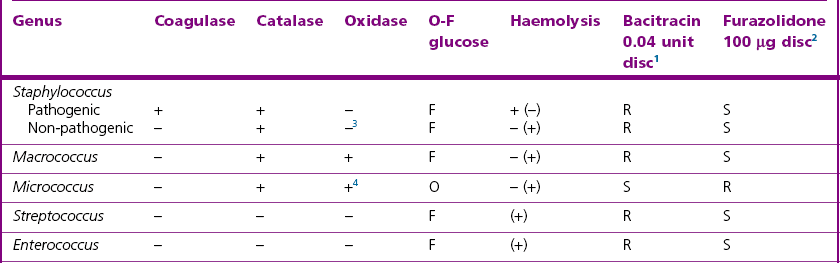
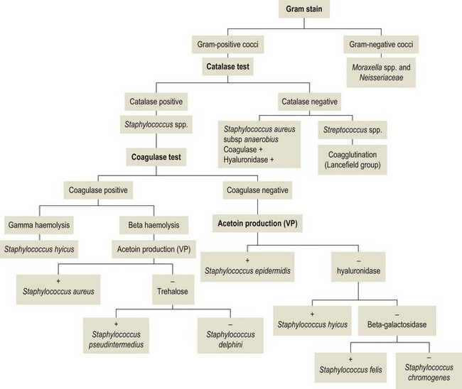
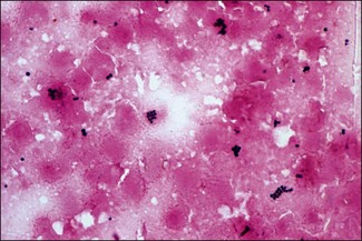
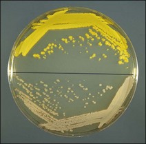
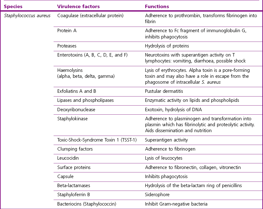
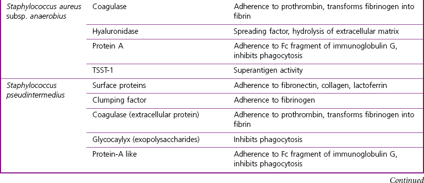
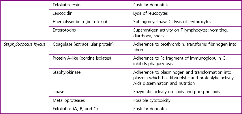
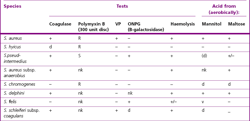
Chapter 7 The staphylococci are Gram-positive cocci with an average diameter of 0.8 to 1 µm, that tend to be arranged in pairs, tetrads, or more often, grouped in irregular clusters or ‘bunches of grapes’ (Fig. 7.1). Colonies are usually white with regular edges. They are non-motile, non-sporulating and most species are facultative anaerobes, with a fermentative metabolism. They are sensitive to lysostaphin (MIC of 12.5 µg/ml) and furazolidone (100 µg/disc) and resistant to lysozyme (MIC of 1000 µg/ml), bacitracin (0.04 unit disc) and to O/129 (0.5 mg). They are usually catalase-positive and oxidase-negative. Growth occurs on nutrient and blood agars but not on MacConkey agar. They are usually not capsulated or have a limited amount of capsule. There are about 30 species of staphylococci and most are found in animals but few are pathogenic. They are considered opportunistic pathogens. Infections with staphylococci are often acute and pyogenic. The two major pathogenic staphylococci, Staphylococcus aureus and S. pseudintermedius are coagulase-positive. The coagulase test usually correlates well with pathogenicity. However, the cause of exudative epidermitis in young pigs, Staphylococcus hyicus, can be coagulase-negative as only 24 to 56% of isolates are coagulase-producing isolates. Coagulase-negative staphylococci occur as commensals and in the environment. They are considered a major component of the normal microflora of animals and humans and occasionally cause opportunistic infections. In this chapter, we will focus primarily on the identification of S. aureus, S. pseudintermedius, S. hyicus, S. chromogenes, S. aureus subsp. anaerobius, S. delphini, S. schleiferi subsp. coagulans and S. felis, the species most commonly associated with animal infections. Micrococci are non-pathogenic, Gram-positive cocci that could be confused with coagulase-negative staphylococci. However, micrococci are variably positive to conventional oxidase tests, oxidase-positive in a modified oxidase test (Faller & Schleifer 1981), are oxidative in the O-F test and have a different susceptibility pattern to bacitracin and furazolidone. The colonies of the micrococci can be white but are often pigmented, the pigmentation ranging from a garish-yellow through cream, to buff or pink (M. roseus) (Fig. 7.2). Streptococci and enterococci are distinguished from staphylococci by the catalase test. Macrococci cells are approximately 4 to 5 times bigger than staphylococcal cells with a diameter of about 2 µm. The pertinent reactions for the commonly isolated Gram-positive cocci are summarized in Table 7.1. Table 7.1 The main differentiating characteristics of the Gram-positive cocci 1Susceptible to bacitracin = zone 10–25 mm 2Susceptible to furazolidone = zone 15–35 mm 3Except Staphylococcus sciuri which is oxidase positive Enzymes include staphylokinase which is a plasminogen activator; coagulase which causes plasma coagulation in vitro; hyaluronidase (‘spreading factor’); lipase; collagenase; proteases; nucleases and urease, all of which may have a role in the pathogenesis of staphylococcal infections. Table 7.2 lists the main diseases caused by the pathogenic staphylococci while their main virulence factors are presented in Table 7.3. Table 7.2 Main diseases caused by the pathogenic staphylococci in veterinary medicine The species characteristics discussed in the paragraphs below are those usually used to identify these organisms in a diagnostic veterinary bacteriology laboratory. Key tests for rapid identification of the most clinically significant Staphylococcus species are found in Table 7.4 while a schematic representation is shown in Figure 7.3. Figure 7.3 Flow chart outlining rapid identification of most clinically significant Staphylococcus species.
Staphylococcus species
Genus Characteristics
Staphylococci Compared with Other Gram-Positive Cocci
Pathogenesis and Pathogenicity
Species
Host(s)
Diseases
Staphylococcus aureus
Many animal species
Abscesses and suppurative conditions. Infection can be systemic. Important cause of infections following surgery
Cattle
Mastitis: subclinical, chronic, acute, peracute or gangrenous
Udder impetigo: small pustules, often at base of teats
Sheep
Mastitis: acute, peracute or gangrenous
Tick pyaemia of lambs (two to five weeks old): associated with heavy tick (Ixodes ricinus) infestation
Periorbital eczema (dermatitis): infections of abrasions, associated with communal trough feeding
Dermatitis: predisposed to by scratches from vegetation such as thistles
Goats
Mastitis: subacute or peracute
Dermatitis
Pigs
Mastitis: acute, subacute and chronic (botryomycosis)
Necrotizing endometritis
Udder impetigo: after abrasions from teeth of piglets
Horses
Mastitis: acute
Botryomycosis (spermatic cord) after castration
Rabbits
Exudative dermatitis in neonates
Abscesses, conjunctivitis and pyaemic conditions
Poultry
‘Bumble-foot’: pyogranulomatous lesion of subcutaneous tissue of foot that can involve the joints
Arthritis and septicaemia in turkeys
Omphalitis (more commonly caused by Escherichia coli)
Dogs, cats
Suppurative conditions similar to those listed for S. pseudintermedius
Staphylococcus aureus subsp. anaerobius
Sheep
Lesions similar to those of caseous lymphadenitis (Corynebacterium pseudotuberculosis)
Staphylococcus pseudintermedius
Dogs, cats
Canine (feline) pyoderma (juvenile and adult). Chronic and recurrent pyoderma is a complex syndrome possibly involving cell-mediated hypersensitivity, endocrine disorders and a genetic predisposition. Responds poorly to antibiotic therapy alone
Pustular dermatitis occurs in neonates or in adults under conditions of poor hygiene. Responds readily to antibiotic therapy
Pyometra
Otitis externa (usually in concert with other pathogens)
Infections involving respiratory tract, bones, joints, wounds, eyelids and conjunctiva
Horses, cattle
Rare infections in these species
Staphylococcus hyicus
Pigs
Exudative epidermitis (greasy pig disease), usually in pigs under seven weeks old, there is systemic involvement and the condition can be fatal
Septic polyarthritis, metritis, vaginitis
Cattle
Rare cases of mastitis and cutaneous infections
Horses
Cutaneous infections
Staphylococcus chromogenes
Ruminants
Pigs
Horses, cats
Subclinical mastitis
Exudative epidermitis
Dermatitis (rare)
Staphylococcus delphini
Dolphins
Purulent cutaneous lesions
Staphylococcus felis
Cats
Otitis, abscesses, dermatitis, cystitis, conjunctivitis
Staphylococcus schleiferi subsp. coagulans
Dogs
Otitis externa
Species Characteristics
< div class='tao-gold-member'>
![]()
Stay updated, free articles. Join our Telegram channel

Full access? Get Clinical Tree


Staphylococcus species
Only gold members can continue reading. Log In or Register to continue
