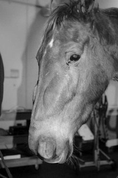
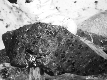
Jeffrey Phillips Splenic tumors are rare in horses, accounting for less than 1% of all diagnosed tumors. Splenic tumors include both benign and malignant variants and may constitute either primary or metastatic lesions from other primary tumor sites, with metastatic being the most common. These tumors may arise from the vascular endothelium (angio-), vascular smooth muscle (leiomyo-), connective tissue component (fibro-), and hemolymphatic tissues. Although no true case series has reported the prevalence for each of these different subtypes of splenic neoplasia, the most common types diagnosed clinically are the lymphoproliferative diseases, including lymphoma and leukemia. Few can forget the great U.S. Champion Sprint Horse Lost in the Fog who succumbed to lymphoma in 2006. Metastatic tumors of the spleen have been described from necropsy surveys of abdominal neoplasia in the horse. The most common metastatic tumors are gastrointestinal tract (gastric ≫ small intestinal > colonic) carcinomas. Other, less common metastatic tumors include gastrointestinal stromal cell tumors, renal cell carcinomas, ovarian carcinomas, and uterine carcinomas and sarcomas. Metastatic tumors of the spleen originating from primary sites outside the abdominal cavity have also been described, with the most common being metastatic melanoma (Figure 100-1). Because benign tumors of the spleen are typically not diagnosed clinically, they will not be reviewed here. Malignant tumors that arise from the mesenchymal component include fibrosarcoma, leiomyosarcoma, and hemangiosarcoma. At the time of diagnosis, these tumors are typically locoregionally advanced or widely metastatic, which limits therapeutic options. Round cell tumors of the spleen, including histiocytic sarcoma and lymphoid malignancies, have been reported in the horse. Histiocytic sarcoma has been described as part of a widely disseminated process and is typically referred to as malignant histiocytosis. Lymphoma of the spleen is also typically diagnosed as a part of a multicentric disease process but may arise as isolated splenic disease. Lymphoid tumors of the spleen are typically high grade and diffuse, with a large non–cleaved cell morphology, although small cell lymphomas of the spleen have also been described. The growth patterns of splenic lymphoma include both solitary mass lesions and diffuse infiltrative growth. Most cases of splenic lymphoma (>80%) are B-cell malignancies that are positive for the BLA-36 antigen. Approximately one-third of these cases are further classified as T-cell–rich large B-cell lymphoma variants. In those variants, the neoplastic population consists of BLA-36–positive large B cells, whereas the CD3+ T cells appear to be a nonneoplastic reactive component. Diagnosis of primary splenic neoplasia is generally accomplished with a combination of transabdominal ultrasound and fine-needle aspiration with cytologic evaluation of tumorous lesions. The ultrasonographic appearance of splenic lymphoma in particular includes solitary or diffuse hypoechoic masses to nodules, but diffuse hyperechoic changes have also been described. Ultrasound-guided biopsy or aspirates of splenic lesions are routine, although peritoneal fluid analysis has also proved useful for diagnosis of intraabdominal neoplasia, including splenic tumors. Hemorrhagic effusions are common in horses with hemangiosarcoma, but neoplastic cells are unlikely to be identified within the fluid. In fact, solid malignant tumors such as sarcomas and carcinomas rarely exfoliate adequately to yield diagnostic samples, whereas lymphoid tumors and metastatic melanoma exfoliate well and can routinely be diagnosed from abdominal effusions. Serologic assays (immunoglobulin M analysis) and blood work changes have also been described in horses with abdominal neoplasia, but these changes are not pathognomonic for splenic tumors. After the diagnosis of primary splenic neoplasia, treatment options depend on tumor type and extent of disease. Diagnostic tests useful for determining the extent of disease include complete blood work, thoracic radiographs, abdominal ultrasound, peritoneal fluid analysis, and rectal palpation. Use of laparoscopy in diagnosis of both benign and malignant splenic tumors has been reported and may also be helpful in evaluating for intraabdominal spread. In horses with isolated solid tumors or even in rare cases of splenic lymphoma, the treatment of choice is splenectomy. Several approaches have been described for splenectomy, and most necessitate rib resections for adequate exposure. Transthoracic approaches, from the 16th rib cranially, also involve incision of the diaphragm, whereas a purely transabdominal approach can be accomplished by resection of the 17th rib. Laparoscopy-assisted abdominal approaches have also been used successfully for splenectomies, reducing both incision size and overall morbidity. For horses with lymphoid malignancies that are not candidates for primary splenectomy, chemotherapy may be attempted for disease palliation. Several chemotherapy regimens have been used in equine patients and include varying doses of prednisolone, vincristine, cyclophosphamide, and doxorubicin (Table 100-1). The effectiveness of these drugs either singly or in combination is unknown but is generally thought to be low. TABLE 100-1 Drugs and Dosages Used in Horses for Treatment of Localized and Systemic Tumors* * Cyclophosphamide, vincristine, and doxorubicin cause severe tissue damage if injected subcutaneously and are for IV administration only. When calculating maximum dosages, the average horse has a body surface area of approximately 5 to 6 m2, resulting in the maximum dosages listed. Maximum dosage for Regressin-V has not been reported. Primary splenic tumors in the horse are rare, whereas metastatic tumors are relatively common, with melanoma being the most common metastatic lesion of the spleen. The biologic behavior of primary splenic tumors is unknown but is generally considered aggressive. For example, hemangiosarcoma and lymphoma of the spleen are commonly described in the literature as widespread diseases at the time of diagnosis. Nevertheless, in horses with solitary tumors, splenectomy may be considered. The prognosis for horses treated with splenectomy for primary tumors of the spleen depends on the histologic nature of the tumor and disease extent; however, long-term survival has been reported for both benign and malignant variants. Tumors of the soft tissues include benign and malignant variants and are commonly diagnosed in horses, comprising approximately 5% of all tumors. For the purposes of this chapter, tumors with primary epidermal involvement (e.g., sarcoids and squamous cell carcinoma) will not be considered here. Malignant soft tissue tumors include both primary and metastatic lesions, although true metastatic lesions in the soft tissues are rare and likely include only melanoma and lymphoma. Primary soft tissue tumors arise from the subcutis and deeper tissues, including muscle, vasculature, and connective tissues. The most common of the malignant primary soft tissue tumors are sarcomas and are typically classified as soft tissue sarcomas, whereas the most common benign soft tissue tumors are fibromas. These benign and malignant variants together make up more than 95% of all soft tissue neoplastic lesions diagnosed in horses. There appears to be no sex predilection for development of soft tissue tumors, although tumor development has been associated with chronic inflammation and prior presence of sarcoid lesions. Ultraviolet light exposure does not appear to be associated with soft tissue tumor formation. Benign tumors of the soft tissues typically arise from either spindyloid-type cells (fibromas) or adipose tissue (lipomas). Fibromas most commonly develop in the bony tissues of the oral cavity or in the nasal or paranasal sinuses but are rarely reported in appendicular locations (Figure 100-2). In contrast, lipomas are generally found internally within the mesenteric adipose tissue but may occasionally be seen at external locations. Benign tumors grow by expansion, resulting in compression and local mass effects on surrounding tissues. Malignant tumors of the soft tissue are also generally composed of spindyloid-type cells and classified as fibrosarcomas or nerve sheath tumors. These so-called fibroblastic sarcomas are clinically considered more aggressive variants of sarcoids. Other histologic types commonly diagnosed include angiosarcoma (hemangiosarcoma, lymphangiosarcoma), undifferentiated sarcoma, histiocytic sarcoma, and leiomyosarcoma; extraskeletal osteosarcoma, giant cell tumor, and other subtypes are more rarely seen. Histologic grading systems have not been developed for equine soft tissue malignancies, but vascular invasion, increased mitotic index, and significant tumor necrosis are clinically associated with more aggressive behavior.
Splenic and Other Soft Tissue Tumors
Splenic Tumors
Histologic Types
Diagnosis and Treatment Options
Drug
Dosage
Route
Cisplatin
1 mg/cm3 of tumor
Intratumoral only; max. ~100 mg per 1000 lb
Carboplatin
225 mg/m2
10 mg/cm3 of tumor
IV
Intratumoral max. ~1200 mg per 1000 lb
5-Fluorouracil
50 mg/cm3 of tumor
Intratumoral max. ~800 mg per 1000 lb
Cyclophosphamide
200 mg/m2
IV only
Vincristine
0.5 mg/m2
IV only
Doxorubicin
35 mg/m2
IV only
Regressin-V
~0.25 mL/cm3 of tumor
Intratumoral only
Summary
Tumors of the Soft Tissues
Histologic Types
< div class='tao-gold-member'>
![]()
Stay updated, free articles. Join our Telegram channel

Full access? Get Clinical Tree


Splenic and Other Soft Tissue Tumors
Chapter 100
Only gold members can continue reading. Log In or Register to continue
