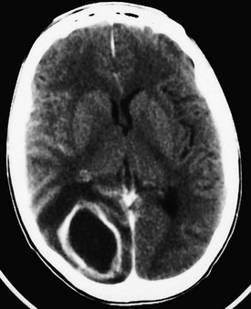
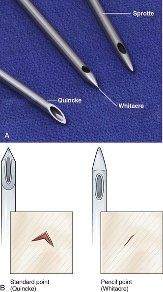
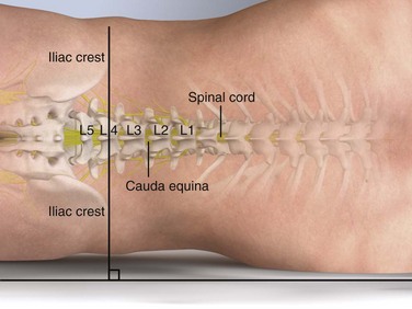
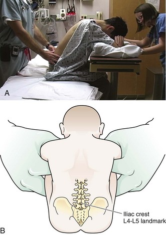
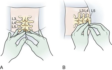
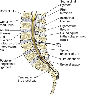
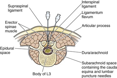
Chapter 60 In 1885, Corning punctured the subarachnoid space to induce cocaine anesthesia in a living patient.1 In 1891, Quincke first removed CSF in a diagnostic study and introduced the use of a stylet.2 He studied the cellular contents and measured protein and glucose levels. Quincke was also the first to record CSF pressure with a manometer. Subsequently, increasingly sophisticated bacteriologic, biochemical, cytologic, and serologic techniques were introduced. In 1919 Dandy reported on replacing CSF with air to determine normal brain anatomy and to identify pathologic changes.3 Water-soluble contrast media have since been used to delineate the spinal subarachnoid space and cerebral cisterns. Other uses of spinal dural puncture include drainage of fluids and injection of anesthetic agents, chemotherapeutic agents, and antibiotics. CSF may have an embryologic nutritive function; at maturity, CSF most likely acts as a mechanical barrier between the soft brain and the rigid fibroosseous dura, skull, and vertebral column. It also appears to support the weight of the brain.4 When buoyed by CSF, the functional weight of the brain is reduced from 1400 to 50 g. Contraction and expansion of CSF accommodates changes in brain volume. The indications for spinal puncture have been reduced with the introduction of noninvasive diagnostic procedures—magnetic resonance imaging (MRI) and computed tomography (CT). A few clinical situations require early or even emergency spinal puncture. The primary indication for an emergency spinal tap is the possibility of central nervous system (CNS) infection (meningitis), with the exception of a suspected brain abscess or a parameningeal process. The need for early detection of meningitis results in the performance of many more lumbar punctures than ultimate diagnoses of infection.5 No other method can be used to completely exclude meningitis. The mere presence of a fever does not mandate lumbar puncture. However, CSF should generally be examined for evidence of infection in patients with a fever of unknown origin, especially if consciousness is altered or the immune system is impaired, even in the absence of meningeal irritation. Meningeal signs may not be present in patients who are old, debilitated, or immunosuppressed; are receiving antiinflammatory drugs; or have had partial treatment with antibiotics.6,7 In a newborn, even a fever is not a dependable sign because temperatures may be normal or even subnormal. For infants younger than 1 year, a high index of suspicion is required to make the diagnosis of meningitis. Approximately 25% of infants with meningitis will not have nuchal rigidity, but many appear toxic or moribund. A tense and bulging fontanelle is somewhat more reliable, although this sign may be absent in a dehydrated child. Neonatal meningitis occurs in 25% of sepsis cases. In addition, 15% to 20% of infants with meningitis have negative blood cultures.8,9 In a child between the ages of 1 month and 3 years, fever, irritability, and vomiting are the most common signs of meningitis. Typically, handling is painful for the child, and the child cannot be comforted. In addition, an older child may complain of a headache. At all ages, patients generally appear ill and drowsy with a dulled sensorium. Physical signs become more useful in diagnosing meningitis in children older than 3 years10 and include nuchal rigidity, Kernig’s sign (effort to extend the knee is resisted), and Brudzinski’s sign (passive flexion of one hip causes the other leg to rise, and effort to flex the neck makes the knees come up). The jolt accentuation test is a more sensitive sign of meningeal irritation.11 This test is considered positive when the patient’s pain is exacerbated by lateral rotation of the head to either side. A petechial rash in a febrile patient should also raise suspicion for Neisseria meningitis.12 Previous use of antimicrobial agents may modify the clinical and CSF findings; partially treated children are less likely to be febrile or exhibit an altered mental status. In addition, patients in the early stages of meningitis may lack the classic features associated with advanced disease. The second indication for emergency spinal puncture is suspected spontaneous subarachnoid hemorrhage (SAH). The diagnosis is usually made by imaging (head CT) or by the direct finding of blood in CSF obtained by spinal puncture.13,14 The sensitivity of CT has been estimated to be as high as 95% soon after hemorrhage, but it drops in patients evaluated later than 24 hours after the event to 76% after 48 hours and to about 50% at 1 week. Before a major hemorrhage, 20% to 60% of patients with aneurysmal SAH have had a “sentinel thunderclap” or “warning leak” headache (i.e., an unusual sudden headache caused by a “minor” leak of blood from the aneurysm). The headache may precede a major rupture by hours to months. The goal of early clinical recognition is surgical or other interventional therapy before a recurrent major hemorrhage. After a warning leak, head CT is more likely to be negative, thus giving added importance to the performance of a lumbar puncture. Migraine or “vascular” headache is a common misdiagnosis in patients with SAH who initially have negative findings on head CT. The usual clinical picture of SAH is an instantaneous excruciating headache. Patients generally recall the exact moment that the headache occurred. The location of the headache is variable and does not necessarily indicate the site of hemorrhage. Nausea, vomiting, and prostration are common symptoms, with approximately one third of patients becoming unconscious at the onset. Examination reveals an acutely ill patient with irritability or overt altered mental status. Meningeal signs are commonly present at the time of initial examination and usually develop within 2 to 3 days. The meningeal signs may become more severe during the first week after hemorrhage and correspond to the breakdown of blood in CSF. During the first week many patients are febrile, a reflection of chemical hemic meningitis.15 Failure to detect blood radiographically in an awake patient may indicate a small hemorrhage or a predominant basal accumulation of blood. If a patient is initially seen several days after the hemorrhage, the blood may have become isodense with brain tissue and may no longer be visible on CT. The proper diagnosis would then require spinal puncture. Newer CT scanners detect recent SAH quite accurately.16 However, because acute SAH is not detected by the initial CT scan in at least 2% to 5% of all patients, lumbar puncture is appropriate to rule out the diagnosis with certainty.17–19 If the neurologic picture demonstrates localizing findings, the presence of a large intracranial hematoma should be suspected, and spinal puncture is contraindicated until imaging studies delineate the nature of the lesion. Idiopathic intracranial hypertension (IIH) is a rare condition of unclear etiology. Patients with IIH have a marked elevation in intracranial pressure (ICP; usually to 250 to 450 cm H2O) without hydrocephalus or mass lesions in the setting of normal CSF composition. Findings on neuroimaging studies are typically normal, and most cases are idiopathic. Because of the nonspecific signs and symptoms, many cases initially escape detection by clinicians. IIH has been associated with hypervitaminosis A, tetracycline, estrogen therapy, and a plethora of other conditions such as sarcoidosis, tuberculosis, and carcinomatosis. Because of the nonspecific findings, many cases initially escape detection by clinicians. IIH is most common in obese adolescent girls and young women, but it can also occur in children and men. Patients suffer from chronic headaches, typically worse with maneuvers that increase ICP (e.g., Valsalva maneuver, squatting, bending, coughing). Papilledema is frequently present. Cranial nerve palsies, particularly of the sixth cranial nerve, are due to increased ICP alone and may be falsely localizing.20 Spinal puncture is absolutely contraindicated in patients with infection in the tissues near the puncture site.4,21 Spinal puncture is relatively contraindicated in the presence of increased ICP caused by a space-occupying lesion. Caution is particularly advised when lateralizing signs (hemiparesis) or signs of uncal herniation (unilateral third-nerve palsy with an altered level of consciousness) are present. In such cases, a tentorial or cerebellar pressure cone may be precipitated or aggravated by the spinal puncture. Cardiorespiratory collapse, stupor, seizures, and sudden death may occur when pressure is reduced in the spinal canal.22 The risk for herniation seems to be particularly pronounced in patients with brain abscesses.23,24 Brain abscesses are frequently manifested as expanding intracranial lesions that induce headache, mental disturbances, and focal neurologic signs rather than as an infectious process with signs of meningeal irritation. Fever is often absent. In 75% of cases, a primary source of chronic suppuration is present. Common predisposing factors for brain abscess include craniofacial trauma; craniocerebral trauma; penetrating injuries that push bone fragments into the brain; large animal bites of infants’ skulls; neurosurgical procedures; cardiovascular disorders treated with right-to-left shunts; bacterial endocarditis; gram-negative sepsis in neonates; dental infections; chronic sinusitis; otitis; mastoiditis; chronic abdominal, pulmonary, or pelvic infection; bacterial meningitis; and immunosuppression. Abscesses may also develop in infarcted brain tissue in septic patients if the blood-brain barrier is compromised.25 Although the CSF is usually abnormal (elevated pressure, elevated white blood cell [WBC] count, and elevated protein concentration), spinal puncture in patients with a known or suspected abscess is contraindicated. Brain herniation markedly reduces the patient’s likelihood of survival. If the history suggests abscess, CT can rapidly diagnose and localize the lesion (Fig. 60-1).26 Because the appearance of brain abscesses on CT is similar to that of neoplastic and vascular lesions, false-positive reports of brain abscess are possible.26 Spinal epidural hematomas are very rare but may occur in certain subpopulations of patients undergoing lumbar puncture. Those most at risk are individuals with a bleeding diathesis, including those treated with anticoagulants either before or immediately after lumbar puncture and those with abnormal clotting mechanisms, especially thrombocytopenia. The condition can rarely occur even with a nontraumatic tap and in the absence of a coagulation defect.27 Spinal subdural hematomas after lumbar puncture are even rarer.28,29 Lumbar puncture can injure the dural or arachnoid vessels, which may result in minor hemorrhage into the CSF. This is generally of little consequence. However, the number of patients with hemophilia and human immunodeficiency virus (HIV) infection who require lumbar puncture has increased since the late 1990s. In a coagulopathic patient, attempt to correct the clotting deficiency if clinically feasible and time permits. The procedure should be performed by experienced clinicians, who are less likely to traumatize the dura. After the procedure, monitor the patient carefully for progressive back pain, lower extremity motor and sensory deficits, and sphincter impairment. Thoroughly investigate any complaints of motor weakness, sensory loss, or incontinence after lumbar puncture. Lumbar puncture may be performed in patients with a coagulation defect if the procedure is expected to provide essential information such as in the diagnosis of meningitis and the pathogen responsible. In cases of severe thrombocytopenia, infusion of platelets before the puncture may be desirable. Correct warfarin-induced coagulopathy with fresh frozen plasma (FFP) or prothrombin complex concentrate together with vitamin K. However, the vitamin K–FFP protocol may take 12 to 24 hours to totally reverse the effect of warfarin. Correct the coagulopathy before a puncture if the clinical situation permits such delay. Lumbar puncture can be performed safely in patients with hemophilia A or B whose deficient clotting factor is replenished before the procedure.30 Additional replacement of the clotting factor after the procedure is of unknown value. Performance of lumbar puncture in patients with leukemia and low platelet counts has also been studied. Howard and coworkers reported on 5223 lumbar punctures performed on 958 children with newly diagnosed acute lymphoblastic leukemia.31 The platelet count was 10 × 109/L or lower in 29 children, 11 to 20 × 109/L in 170 children, and 21 to 50 × 109/L in 742 children. No serious complications were reported in any group. The overall rate of traumatic taps was 10.5%, but such taps were not associated with adverse sequelae. The authors concluded that in children with acute lymphoblastic leukemia, prophylactic platelet transfusion for lumbar puncture is not required if the platelet count is higher than 10 × 109/L. The number of patients with platelet counts lower than 10 × 109/L was too small to allow any conclusion about this group. A more recent review concluded that in the absence of additional risk factors, a platelet count of 40 × 109/L is “safe” for lumbar puncture. Lower counts are probably safe as well, but there was insufficient evidence to make recommendations.32 Assemble the standard equipment for a spinal puncture before starting the procedure. Place it where the operator can easily access it. A standard-point Quincke cutting needle is most often used and supplied with the kit. Some operators prefer to use a Sprotte needle (Havel’s, Inc., Cincinnati, OH) or a Whitacre needle (Becton Dickinson and Company, Rutherford, NJ) to minimize any dural injury associated with passage of the needle (Fig. 60-2). These styletted needles have a side port for withdrawal of fluid and, theoretically, are more likely to separate than cut the dural tissue. Although commercial kits provide most of the items needed for lumbar puncture, the operator should bring additional supplies, including supplemental spinal needles, specimen tubes, gauze, antiseptic solution, additional local anesthetic, needles and syringes, and extra sterile gloves of the appropriate size (see Review Box 60-1). Lumbar puncture is commonly carried out with the patient in the lateral recumbent position (Fig. 60-3). A line connecting the posterior superior iliac crests intersects the midline at approximately the L4 spinous process (Fig. 60-4). Spinal needles entering the subarachnoid space at this point are well below the termination of the spinal cord, and the only important neurologic structure is the cauda equina. Generally, the needle pushes isolated nerves to the side during advancement. The adjacent interspace above or below may be used, depending on which area appears to be most accessible to palpation. The space between the lumbar vertebrae is relatively wide. In the thoracic region, the spinous processes overlap and are directed caudally; therefore, there is no midline area free of overlying bone. In adults, the spinal cord extends to the lower level of L1 or the body of L2 in 31% of persons, thus eliminating higher levels as sites for puncture. Puncture in adults and older children may be performed from the L2-L3 interspace to the L5-S1 interspace. Some clinicians place the patient in an upright sitting position because the midline is more easily identified. This position can be used in both adults and infants (Fig. 60-5). The higher CSF hydrostatic pressure while sitting may aid flow of CSF in a dehydrated patient. Observe caution regarding orthostatic changes in blood pressure and airway maintenance. Generally, allow a sitting patient to lean onto a bedside stand and use a pillow to rest the head and arms. Have an assistant support the patient during the procedure. Radiographic studies by Fisher and colleagues have demonstrated the advantages of hip flexion when the sitting position is used.33 To accomplish hip flexion, use a stool to support the patient’s feet, which pulls the knees up toward the chest. This increases lumbar interspinous width, which may increase the success and ease of needle passage. Iatrogenic infection after lumbar puncture is extremely rare. Use sterile gloves during the procedure, but the need for face masks is debatable.34–36 Apply the same guidelines for control of central line infection (caps, gowns, gloves, and masks) to lumbar puncture.37 Wash the patient’s back with an antiseptic solution applied in a circular motion and increase the circumference of the cleansed area with each motion. Place a sterile towel or drape between the patient’s hip and the bed. Commercial trays have a second sterile drape with a hole that may be centered over the site selected for the procedure. With the patient in the lateral decubitus position, place the needle into the skin in the midline, parallel to the bed. Hold the needle between both thumbs and index fingers (Fig. 60-6). After the subcutaneous tissue has been penetrated, angle the needle toward the umbilicus. The bevel of the needle should be facing straight up toward the ceiling. The supraspinal ligament connects the spinous processes, and the interspinal ligaments join the inferior and superior borders of adjacent spinous processes. The ligamentum flavum is a strong, elastic membrane that may reach a thickness of 1 cm in the lumbar region. The ligamentum flavum covers the interlaminar space between the vertebrae and assists the paraspinous muscles in maintaining an upright posture (Fig. 60-7). The ligaments are stretched in a flexed position and are more easily crossed by the needle. The ligaments offer resistance to the needle, and a “pop” is often felt as they are penetrated. Hold the stylet in place during advancement but remove it frequently to see whether the subarachnoid space has been reached. The pop may not be felt with the very sharp needles contained in disposable trays. If bone is encountered, partially withdraw the needle to subcutaneous tissue. Repalpate the back and ascertain that the needle is in the midline. Directing the tip of the needle toward the navel often enhances navigation of the interspinal space. If bone is encountered again, slightly withdraw and reangle the needle so that the point is placed at a more sharply cephalad angle (Fig. 60-8). This approach should avoid the inferior spinous process. Normal CSF is a clear fluid and will flow from the needle when the subarachnoid space has been penetrated and the stylet is removed. In normal patients, the dura will be penetrated when the needle is advanced about one half to three fourths of its length. In obese patients, the entire length of the needle may be required to reach the subdural space (Fig. 60-9). If feasible, attach the manometer and record the opening pressure (Fig. 60-10). This step is commonly omitted in critically ill patients. Pressure readings from a struggling patient may be inaccurate. Readings are valid only if taken with the patient relaxed and in the lateral decubitus position (not the sitting position). A three-way stopcock, supplied in disposable trays, allows both collection of CSF and measurement of opening pressure with a single needle. Positioning of the manometer is often more convenient if an extension tube (provided with most disposable trays) connects the needle hub to the stopcock, which in turn is attached to the manometer. Position the manometer so that the “zero” mark is at the level of the spinal needle. Then ask the patient to relax. Extending the legs after needle placement does not meaningfully decrease opening CSF pressure.38 The observation of phasic changes in the fluid column with respirations and arterial pulsations confirms needle placement in the subarachnoid space. If the needle is against a nerve root or is only partially within the dura, the pressure may be falsely low, and respiratory excursions will not be reflected in the manometer. Minor rotation of the needle may solve these problems. Hyperventilation will reduce the pressure readings because of hypocapnia and the resultant cerebral vasoconstriction. Traumatic taps are common and usually clinically inconsequential. Nevertheless, they can be minimized by proper patient and needle positioning. A traumatic tap most commonly occurs when the subarachnoid space is transfixed at the entrance of the ventral epidural space, where the venous plexus is heavier. A plexus of veins forms a ring around the cord, and these veins may be entered if the needle is advanced too far ventrally or is directed laterally (Fig. 60-11). If blood is encountered and the fluid does not clear, repeat the procedure at a higher interspace with a fresh needle. A traumatic tap, per se, is not a particularly dangerous problem in a patient with normal coagulation, and no specific precautions are needed if blood-tinged fluid is obtained. However, observe for signs of cord or spinal nerve compression from a hematoma developing within the first several hours in patients with a coagulopathy. The supraspinal ligament might be calcified in older persons, thus making a midline perforation difficult. A calcified ligament may deflect the needle. In this case, use a slightly lateral approach. Because the lower lamina rises upward from the midline, direct the needle slightly cephalad to miss the lamina and slightly medially to compensate for the lateral approach. The needle passes through the skin, superficial fascia, fat, the dense posterior layer of the thoracolumbar fascia, and the erector spinae muscles. The needle then penetrates the ligamentum flavum (bypassing the supraspinal and interspinal ligaments), the epidural space, and the dura before CSF is obtained (Fig. 60-12). Lateral cervical puncture is an alternative approach that can be performed utilizing an insertion site 1 cm inferior and 1 cm dorsal to the mastoid process.39 Lumbar puncture in infants is usually performed to exclude meningitis or encephalitis. The sitting position may allow the midline to be identified more easily. Use of a needle without a stylet has been suggested for small infants because this device allows the pressure to be estimated as the needle punctures the dura.40 However, failure to use a stylet may cause the subsequent development of an intraspinal epidermoid tumor.41 Use of a butterfly infusion set needle simplifies the procedure, which is helpful when managing a squirming or hyperactive patient.40 In general, a stylet is recommended primarily at the time of skin penetration and on needle withdrawal, although many operators use the stylet during any advancement of the needle. If the child’s neck is very tightly flexed, CSF might not be obtained. However, if the head is held in midflexion, CSF usually flows briskly. If CSF fails to flow, gently suction with a 1.0-mL syringe to exclude a low-pressure syndrome. Because pressure readings are inaccurate in a struggling child, measurement of pressure is not commonly attempted in infants and young children.42 Avoid prolonged severe flexion of the neck in an infant because it may produce dangerous airway obstruction. If the infant suddenly stops crying, check the airway immediately.43 Proper positioning is best accomplished by an assistant, who maintains the spine maximally flexed by partially overlying the child and using the chest and body weight to immobilize the thorax and hips while holding the child behind the shoulders and knees.44 Infants have poor neck control; therefore, the assistant must also ensure that the child maintains an open airway. Pay particular attention to avoiding marked neck and trunk flexion. Incorrect positioning usually results in multiple punctures and a bloody tap. Newborn and preterm infants may experience significant hypoxia and clinical deterioration during lumbar puncture; a sitting position appears to be preferable.45 Lumbar puncture in infants with respiratory distress syndrome may pose greater risk than benefit.46 This is a problem primarily in neonates but may also apply to younger infants with sepsis. Closely monitor all infants with serious cardiopulmonary disease during the procedure. Preoxygenation with or without monitoring oxygen saturation may be used as a precaution. Although local anesthesia or sedation has not been a routine practice during lumbar puncture in an infant or child, it is being reconsidered.47,48 Neonates perceive pain, and local anesthesia neither produces physiologic instability nor makes the procedure more difficult.49,50 The use of topically applied EMLA (eutectic mixture of local anesthetics) (AstraZeneca) reduces the pain associated with needle insertion in newborns.51 Sedation of an anxious child may be considered, but sedatives are relatively contraindicated in an obtunded patient without a protected airway and in the setting of hemodynamic instability. The traditional approach to lumbar puncture depends on palpation of bony landmarks to determine the correct location for insertion of the needle. However, landmarks are difficult to palpate in overweight and obese patients.52 When lumbar puncture fails, the usual alternative has been fluoroscopic guidance, which necessitates movement of the patient out of the ED, as well as the availability of an appropriately trained radiologist.52 Bedside ultrasound is increasing being used in emergency and critical care medicine, and its scope of practice has expanded to include guidance for a number of procedures, including lumbar puncture (see Ultrasound Box).53–56 Ultrasound can be used in adults, as well as in neonates and infants.57 Lumbar Puncture by Christine Butts, MD The bony spinous process will appear as a hyperechoic crescent-shaped structure with posterior acoustic shadowing. In the transverse view, acquisition of a perfectly symmetric image will provide assurance that the probe is in the anatomic midline (Fig. 60-US1). In the longitudinal view, multiple spinous processes can be visualized, along with the interspaces between them (Fig. 60-US2). To perform ultrasound-guided lumbar puncture, position the patient in the usual manner. Place the ultrasound probe over the spine in a transverse orientation at the level of the iliac crests such that the shadow caused by the spinous process is centered on the screen. Use a pen to mark the skin on each side of the transducer, exactly at the midpoints (Fig. 60-US3). These two marks can then be connected to mark the midline of the spine. Next, rotate the transducer 90 degrees to obtain a midline longitudinal view and position it until the gap between two spinal processes is in the center of the screen. Mark the skin at the midpoint of each side of the transducer and then connect the marks to form a single line (Fig. 60-US4). The intersection of the two lines is the site for entry of the needle (Fig. 60-US5). Cleanse the skin with antiseptic solution and perform the remainder of the procedure in the usual fashion. Figure 60-US3 First use the ultrasound (US) probe in the transverse orientation at the level of the iliac crests, and obtain an image with the shadow of the spinous process centered on the screen (as in Fig. 60-US1). Mark the skin at the exact midpoint of the transducer. This represents the anatomic midline. Figure 60-US4 Rotate the ultrasound (US) probe 90 degrees and obtain a midline longitudinal view. Position the probe so that the interspace is centered on the screen (as in Fig. 60-US2). Mark the skin at the midpoint of the transducer. This represents the level of needle entry. Headache after Lumbar Puncture A number of complications from lumbar puncture have been reported.58 One of the most common is headache, which occurs after 1% to 70% of spinal taps.59–66 In general, the development of postpuncture headache can be neither prognosticated nor prevented. The syndrome most commonly starts within the first 48 hours after the procedure (although a case occurring 12 days after the procedure has been reported)67 and usually lasts for 1 to 2 days (occasionally as long as 14 days). Cases lasting months have been described. The headache usually begins within minutes after the patient arises and characteristically ceases as soon as the patient lies down. The pain is mild to incapacitating and is generally cervical and suboccipital in location but might involve the shoulders and the entire cranium. Exceptional cases include nausea, vomiting, vertigo, blurred vision, ear pressure, tinnitus, and stiff neck. The headache may change to a positional backache or neck ache. The technique of spinal puncture probably has little to do with the development of a postprocedure headache. The syndrome is widely thought to be caused by leakage of fluid through the dural puncture site. This results in a reduction in CSF volume below the cisterna magna and downward movement of brain tissue, along with displacement and stretching of pain-sensitive structures such as the meninges and vessels, and causes a traction headache. The recumbent position brings relief because the weight of the brain is shifted cephalad. Another proposed mechanism is cerebral vasodilation. A more recent hypothesis suggests that the headache is caused by an altered distribution of craniospinal elasticity and acute intracranial venous dilation.68 Some authors have commented on the incidence and severity of post–dural puncture headache as being related to the orientation of the spinal needle bevel and its type and size.69–79 It is clear that the incidence of post–dural puncture headache is lower when the bevel of the needle is oriented parallel to the longitudinal axis of the spine. Until relatively recently the explanation for this was that the parallel bevel would separate rather than cut the longitudinal dural fibers and thus produce a smaller dural hole and less CSF leakage. However, it is now known that the dural fibers are oriented randomly, not longitudinally.80 Even with random fiber orientation, back flexion is more likely to close a dural hole in the longitudinal plane than a hole in the horizontal plane. The two basic types of spinal needles are cutting (Quincke) and noncutting or pencil point (Sprotte and Whitacre). Noncutting needles cause a lower incidence of headache after dural puncture, perhaps because the tip of the needle tends to separate rather than cut the dural fibers. It has traditionally been thought that the dural hole resulting from puncture with a noncutting needle is smaller than that created by puncture with a cutting needle; however, this may not be true. The important difference may be that the cutting needle causes a clean-cut opening in the dura whereas the noncutting needle produces a jagged opening with rough edges.81 The jagged opening may produce a more intense inflammatory response that results in edema and more complete closure of the hole. The use of atraumatic spinal needles is not routine.82 As more spinal kit manufacturers include them in kits, their use might increase.83 A single-institution study suggested that routine use of noncutting needles yields a potential cost savings.84 However, in thick-skinned individuals, passage of a thin, noncutting needle may be technically difficult. Making the initial pass with a thicker cutting needle to the level of the interspinal ligament, followed by removal and advancement of a noncutting needle in the same soft tissue tract, can be helpful. Use of a smaller-diameter needle is one intervention that will probably cause a lower incidence of postpuncture headache because it creates a smaller dural hole. If diameter were the only consideration, as small a needle as possible would be used. However, a needle must provide adequate CSF flow rates to allow timely CSF collection and pressure measurement. With a very small needle, a syringe may be needed to withdraw fluid, and pressure cannot be recorded easily. In addition, technically, a small needle such as a 26 gauge is difficult to place and manipulate into a position in which it does not become intermittently obstructed by nerve roots. When these characteristics are considered, a 20- to 22-gauge atraumatic needle seems to be the best overall choice for diagnostic lumbar puncture.72 Despite common beliefs about techniques and postprocedure interventions, lumbar puncture headaches may not be completely preventable. Studies of the influence of directives such as strict bed rest on postpuncture headache have yielded contradictory results, with no consensus on any specific preventive intervention forthcoming concerning the worsening of, improvement in, or effect on the incidence. Brocker reported a reduction in the incidence of headache from 36.5% to 0.5% when patients lie prone instead of supine for 3 hours after puncture with an 18-gauge needle.85 He postulated that the prone position caused hyperextension of the spine and disrupted alignment of the holes in the dura and the arachnoid, thus making leakage less likely. Thoennissen and coworkers concluded that there was no evidence that longer bed rest after lumbar puncture was better than immediate mobilization or short bed rest in reducing the incidence of headache.86 There appears to be no reason to enforce bed rest following lumbar puncture. Other factors that might influence the incidence of a post–spinal puncture headache were reviewed by Fishman and by Lin and Giederman.21,87 The incidence is higher in young patients than in older patients and is also increased in females and individuals with a history of headache. Many medications have been advocated for the treatment of headache after lumbar puncture: barbiturates, codeine, neostigmine, ergots, diphenhydramine (Benadryl), dimenhydrinate (Dramamine), caffeine, amphetamine sulfate (Benzedrine), ephedrine, intravenous fluids (normal saline, lactated Ringer’s solution), magnesium sulfate, and vitamins.21,88–90 Caffeine is a traditional common initial intervention for postpuncture headache, and it appears to be of benefit.90 Caffeine can be administered in the ED, and pain relief can be seen within a few hours and is thought to result from caffeine’s vasoconstrictive effect on the cerebral vasculature. One suggested regimen is 500 mg of caffeine (sodium benzoate) diluted in 1000 mL of normal saline infused over a 1- to 2-hour period. A second dose can be given if the headache is not relieved. If successful, consumption of caffeinated beverages may be continued as an outpatient. For patients with a prolonged low-pressure headache, placement of an epidural blood patch by experienced operators is highly successful and often provides dramatic relief.91–95 Consider a blood patch for all patients with seriously symptomatic spinal headaches. Perform an epidural tap at the level of the previous lumbar puncture. Use the loss-of-resistance technique with sterile saline to locate the epidural space.96 Draw 10 to 20 mL of autologous blood into a syringe aseptically and slowly inject it (1 to 2 mL every 10 seconds) into the epidural space at the site of the dural puncture.97 Slow or discontinue the injection if back pain or paresthesia develops. Keep the patient supine for 1 hour while hydration is administered intravenously. Relief usually occurs within 20 to 30 minutes after the procedure. Epidural patches are less likely to be effective if symptoms have been present for more than 2 weeks. Pain is relieved when the blood patch forms a gelatinous tamponade that stops the CSF leak and immediately elevates CSF pressure. Patch failures (15% to 20%) are believed to be caused by improper needle placement, injection of an inadequate quantity of blood (after which a second patch is usually successful), or an incorrect diagnosis. Complications reported after placement of an epidural patch include back stiffness, paresthesia, radicular pain, subdural hematoma, adhesive arachnoiditis, and bacterial meningitis.98 The procedure should be used in patients with refractory headaches that do not respond to conservative therapy, and it should be performed by clinicians trained in the procedure.
Spinal Puncture and Cerebrospinal Fluid Examination
Historical Perspective
Anatomy and Physiology
Indications for Spinal Puncture
IIH (Pseudotumor Cerebri)
Contraindications to Spinal Puncture
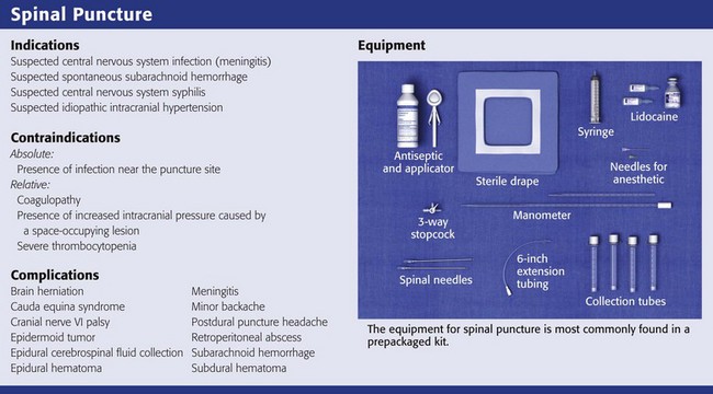
Equipment
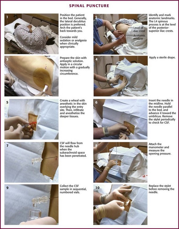
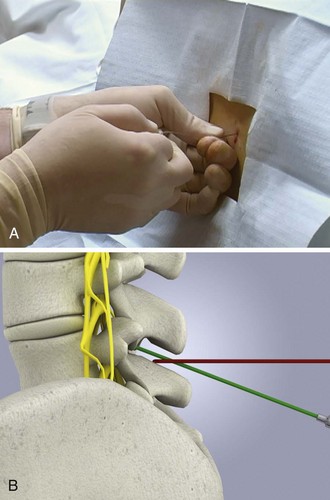
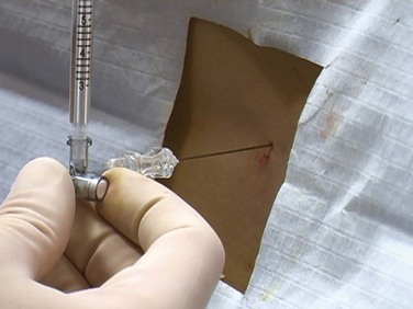
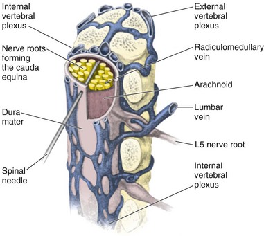
Procedure
Lateral Approach for Lumbar Puncture
Lumbar Puncture in Infants
The Difficult Lumbar Puncture
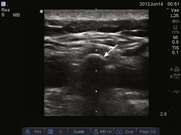
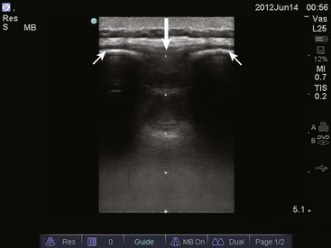
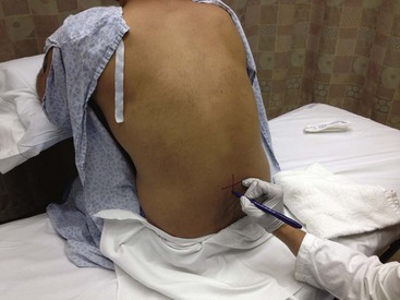
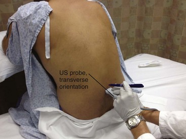
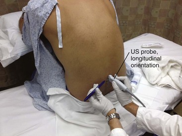
 ULTRASOUND
Complications
![]()
Stay updated, free articles. Join our Telegram channel

Full access? Get Clinical Tree


Veterian Key
Fastest Veterinary Medicine Insight Engine