chapter 18 Special Procedures
Angiocardiography: An intravenous radiographic contrast study evaluating the vascular system and chambers of the heart.
Angiography: An intravenous radiographic contrast study evaluating the vascular system.
Antegrade urethrogram: A method of urethrography in which the contrast medium is voided from the urinary bladder.
Arthrography: A radiographic contrast technique evaluating the articular cartilage, joint space, and joint capsule.
Barium sulfate: A common positive-contrast medium that is available in various forms and is often used as a suspension in gastrointestinal evaluations.
Cholecystography: An oral or intravenous radiographic contrast study evaluating the bile ducts and gallbladder.
Contrast medium: A substance that is either radiolucent or radiopaque and can be administered to increase radiographic contrast within an organ or system.
Cystography: Radiographic contrast studies evaluating the urinary bladder.
Double contrast: A radiographic contrast technique that uses a combination of positive- and negative-contrast media simultaneously.
Double-contrast cystogram: A radiographic study of the urinary bladder involving distending the bladder with a gas and then adding a small amount of positive iodinated contrast medium.
Esophagography: A radiographic contrast study performed to evaluate esophageal function and morphology.
Excretory urography: An intravenous radiographic contrast study of the kidneys and ureters.
Fistula: An abnormal tubelike passage within body tissue.
Fistulography: A positive or negative radiographic contrast study used to determine the depth and origin of a fistulous tract.
Gastrography: A radiographic contrast study performed to evaluate the size, shape, position, and morphology of the stomach.
Intravenous pyelogram (IVP): A radiographic contrast study of the kidney structure and collection system.
Intravenous urogram (IVU): A radiographic contrast study of the kidney structure and collection system.
Lower gastrointestinal (LGI) study: Commonly referred to as a barium enema; a radiographic contrast study evaluating the rectum, colon, and cecum.
Lymphography: A radiographic contrast study evaluating lymphatic vessels and lymph nodes.
Myelography: A radiographic contrast study evaluating the subarachnoid space surrounding the spinal cord.
Negative-contrast agents: Gases that are more radiolucent to x-rays than are soft tissues and have a black appearance on a radiograph.
Nephrogram: A phase of an excretory urogram characterized by the diffuse opacification of the renal parenchyma.
Parasympatholytic agents: Drugs that eliminate the influence of the parasympathetic nervous system.
Pneumocystogram: A negative-contrast radiographic technique evaluating the urinary bladder.
Pneumoperitoneography: A negative-contrast radiographic study consisting of the introduction of a gas into the peritoneal cavity.
Positive-contrast agents: Substances containing elements of high atomic number that are more radiopaque to x-rays than are tissue and bone and have a white appearance on a radiograph.
Positive-contrast cystogram: A radiographic study of the bladder involving distention of the bladder with positive iodinated contrast medium.
Pyelogram: A phase of an excretory urogram characterized by the opacification of the renal collection system.
Retrograde urethrogram: A method of urethrography by which the contrast medium is infused via a catheter placed at the distal end of the urethra.
Sialography: A radiographic contrast study evaluating the salivary glands and ducts.
Triiodinated compounds: A common component of iodinated positive-contrast media that contains three atoms of iodine per molecule.
Upper gastrointestinal (UGI) study: A radiographic contrast study evaluating the stomach and small intestines.
Urethrography: A radiographic contrast study evaluating the urethra.
Vaginography: A radiographic contrast study evaluating the female reproductive organs.
INDICATIONS
Special radiographic procedures are used to supplement or confirm information garnered from routine survey radiographs. Under normal circumstances, soft tissue structures or organs are difficult or impossible to identify on plain films due to lack of contrast. A contrast medium is a substance that is either radiolucent or radiopaque and can be administered to an animal to increase radiographic contrast within an organ or system. With the use of a contrast medium, soft tissue structures can be visualized, and the structure under investigation can be evaluated for size, shape, and position. In addition, defects in the mucosal surface of an organ or its luminal contents can be identified. In some instances it is possible to evaluate organ function or to assess the physiologic condition. Although contrast studies can be extremely helpful for a complete diagnosis, at no time should a special procedure replace routine survey radiography.
CONTRAST MEDIA
Iodine Preparations
Iodine compounds are divided into two subcategories: water-soluble agents and viscous/oily agents.
Oily/viscous agents.
Oily contrast media consist of iodized oils. The oil contains a suspension of propyliodone in either water or arachidic oils. Because of their viscous nature and insolubility in water, they are not resorbed in the body and produce fat embolism. The iodized oils cannot be administered intravascularly. In addition, the agent does not mix with cerebrospinal fluid during myelography. The oils tend to coagulate within the spinal canal and fail to outline lesions clearly. Current practice does not include oily media for myelography. If the agent is not removed, the absorption rate within the spinal canal is extremely slow. The absorption rate is estimated at approximately 1 mL/year.
CONTRAST STUDIES OF THE GASTROINTESTINAL TRACT
Esophagography
Esophagography is performed to evaluate esophageal function and morphology. An esophagogram is indicated for patients with a history of regurgitation of undigested food, acute gagging, or dysphagia. This study consists of administering a positive-contrast medium orally and exposing a number of radiographs during and after the patient swallows the contrast agent. Liquid barium sulfate is usually the contrast medium of choice. Barium sulfate is also available in a thick paste form, which is more difficult to swallow but provides good mucosal coating of the esophagus. Barium can be mixed with canned or hard food, or both, to evaluate the function of the esophagus or for a partial obstruction that may be missed during a plain liquid barium swallow.
Upper Gastrointestinal Study
Precautions.
If the patient is suspected of having a gastrointestinal perforation, barium sulfate is contraindicated. If barium were to enter the abdominal cavity, it would not be absorbed and could induce granuloma formation. In the instance of a perforation, an oral iodinated contrast medium should be used. Unfortunately, iodinated contrast media do not produce as much radiographic contrast. Iodine compounds tend to become diluted as they pass through the bowel because they draw extracellular fluid from the digestive tract. In addition, because of their osmotic activity, they are not recommended for dehydrated patients.
70% to 100% barium sulfate (liquid and paste) or iodinated oral contrast agent
30% to 60% liquid barium sulfate or iodinated oral contrast agent
Enema if necessary 2 to 4 hours before study
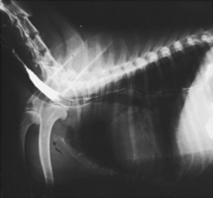
Figure 18-2 Lateral view of an upper gastrointestinal study exposed 5 minutes after the administration of barium.