Chapter 25 Soft Tissue Surgery
Because these herbivorous rodents eat frequently, a short fast of 1 to 2 hours is generally recommended, only to allow the animal to clear its mouth of food material. Herbivores are physiologically unable to vomit, so the risk of aspiration pneumonia is negligible. Because normal gastrointestinal function is vital to recovery, a long fast is not recommended. It has been shown that small mammals with a negative energy balance are at greater risk for postoperative complications.19
The reported total blood volume of small mammals is 57 mL/kg body weight.15,19 With loss of 15% to 20% of the total blood volume, most mammals experience hypovolemic shock and release high levels of catecholamines. Life-threatening consequences usually occur with loss of 20% to 30% of the total blood volume.15,19 This would be equivalent to only 4.5 to 6.8 mL of blood in a 400-g guinea pig. Crystalloid, colloid, whole blood from a conspecific, or a blood substitute can be used in patients experiencing serious blood loss.
Guinea pigs, chinchillas, and degus seem to be less likely to bother surgical incisions than are other species of rodents. In selecting suture material, keep in mind the propensity of these animals to develop a caseous, suppurative response to foreign materials such as sutures. Catgut is degraded by proteolysis and should not be used in rodents because of its reactive nature. Absorbable materials degraded by hydrolysis rather than proteolysis are recommended (e.g., polyglactin 910, polyglycolic acid, polydioxanone, poliglecaprone, glycomer 631, braided lactomer 9-1). Soft, absorbable, braided materials (e.g., polyglycolic acid, polyglactin 910, braided lactomer 9-1) are rapidly absorbed and are less irritating to subcutaneous tissues than stiffer materials such as monofilament absorbable sutures (e.g., polydioxanone, poliglecaprone, glycomer 631). Some surgeons recommend stainless steel for cutaneous sutures; however, this material is very stiff and the cut ends often cause irritation, actually stimulating rather than preventing self-mutilation. Many rodents chew out even steel skin sutures. Skin closure is best accomplished with an intradermal or subcuticular technique. This can be time-consuming if the surgeon is not adept. Skin staples are quickly applied, and most rodents will not bother them because there are no ends to poke and irritate the adjacent skin. Cyanoacrylate tissue adhesive can be used to close small incisions; however, excessive amounts of the adhesive on the skin surface will often attract the attention of the rodent and it will try to groom it off, potentially resulting in dehiscence.
Guinea Pigs, Chinchillas, and Degus
Ovariectomy
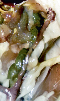
Cystic ovaries are common in guinea pigs, with an incidence reported to be as high as 100%.33,34,37 In a study of guinea pigs with reproductive problems, 76% had cystic ovaries.21 In another study of 43 guinea pigs from various owners or breeders, 53% had evidence of cystic ovaries on ultrasound examination and 36% had evidence of bilateral cystic ovaries.28 Of the guinea pigs above 2 years of age, 93% had cystic ovaries, 62% of which were bilateral. There was a direct relationship between age and the size of the ovarian cysts and there was no difference in incidence between breeding and nonbreeding sows.28 There was no difference in reproductive success in sows with or without cysts younger than 15 months of age; however, guinea pigs greater than 15 months of age with cysts had a marked decrease in reproductive success.21 While cystic ovaries are common, most sows show little evidence of clinical disease. In juvenile sows, they are small (5 μm), but they enlarge as the animal matures, reaching up to 7 cm in size.21,33 If the cysts are <5 mm, there are usually multiple cysts; if they are >2 cm, they are usually single cysts.34 The largest are often multilocular.
Histologically, ovarian cysts in guinea pigs are similar to those of humans and cats, being cysts of the rete ovarii.21,33,34,37 Rete cells are from the mesonephros; they migrate into the fetal gonads and differentiate into rete testis in males and rete ovarii in females. They form a tubular, blind-ended vestigial structure within the ovary. The function of the rete cells is suspected to be phagocytosis of degenerating oocysts and production of a meiosis-inducing substance. These cells do not produce hormones. The pathogenesis of cyst formation is unknown but is suspected to be the result of a defect in ion pumps, so electrolytes are transported into the tubular structure but not out. Fluid is pulled in as the ion concentration increases, resulting in the formation of cysts.
Since these cysts do not produce hormones, patients are usually asymptomatic. Clinical signs include abdominal distention as the cysts enlarge; they may cause anorexia and depression because of the effects of the space-occupying mass. Many sows with ovarian cysts also have uterine disease and may present with a hemorrhagic vaginal discharge or reported hematuria that is actually a result of uterine hemorrhage.9,21 Some guinea pigs have a nonpruritic symmetric alopecia typical of an endocrine alopecia; they become aggressive and begin mounting cage mates9; however, it is considered normal for female guinea pigs to mount cage mates during estrus. These signs are indicative of hormone increases that are difficult to explain, since the cysts do not produce hormones. The diagnosis is confirmed with ultrasound.3 Radiographically, ovarian cysts cannot be differentiated from other abdominal masses because fluid is of the same radiographic density as soft tissues.
Ovariohysterectomy is the treatment of choice for guinea pigs with cystic ovaries because uterine disease secondary to these ovarian cysts is common, although a mechanism has not been established. In a study of five guinea pigs with cystic ovaries histologically confirmed to be rete ovarii cysts, all were found to have uterine disease, including endometritis, pyometra, endometrial hyperplasia, and leiomyomas.9,10
If the uterine changes are secondary to the ovarian cysts, ovariectomy at a young age would be expected to prevent both problems.37 Because ovarian rete cysts are so common and become larger with age, routine ovariectomy or ovariohysterectomy at a young age should be recommended to owners of female guinea pigs. Drainage of ovarian cysts in guinea pigs provides temporary relief, but fluid quickly reforms. Prior to surgery, drain the cysts as well as possible, using percutaneous centesis to decrease their size. Be aware that there are often adhesions between the diseased ovaries and uterus to the body wall and other viscera. In guinea pigs with alopecia, hair regrowth is generally complete within 3 months after surgery.3,9
Rete ovarii cysts do not produce hormones and should not be affected by the administration of gonadotropin-releasing hormone or its analogs, such as human chorionic gonadotropin.5,13,37 Those hormones are effective in treating follicular cysts, causing them to luteinize. Reports of cysts other than cystic rete ovarii are rare, but a granulosa cell tumor was successfully managed by ovariohysterectomy in a guinea pig.5 Still, administration of 100 IU (1,000 USP units) of human chorionic gonadotropin administered intramuscularly in two doses given 2 weeks apart has been reported to resolve the clinical signs temporarily.5 Gonadotropin-releasing hormone has also been reported to be “very effective” in treating guinea pigs with ovarian cysts.25
Ovarian teratomas have also been reported to occur commonly in sows older than 3 years of age.8,11,13 Some teratomas may be as large as 10 cm in diameter.40 They are usually unilateral and rarely metastasize. Affected sows present for depression, weakness, or collapse due to spontaneous intra-abdominal hemorrhage from the tumor. Acute death from blood loss can occur. Ovariectomy or ovariohysterectomy is the treatment of choice for sows with these tumors.
Ovariectomy can be done through a ventral midline ap‑proach or a dorsolateral approach.35 The ventral midline approach is as described for other rodents (see Chapter 28). To perform an ovariectomy using the dorsolateral approach, make a 1- to 2-cm incision on each side ventral to the erector spinae muscle and about 1 cm caudal to the last rib. Bluntly penetrate the muscle with a hemostat and enlarge the opening to about 1 cm. With pressure on the abdomen to push the ovary to the incision, reach in with forceps and grasp the ovary. Exteriorize the ovary and use a hemostatic clip or ligature to control any hemorrhage from the ovarian vessels that might occur. Also be sure to remove the entire oviduct surrounding the ovary to prevent cysts from forming. Close the opening in the body wall with 1 or 2 sutures of monofilament absorbable material and appose the skin edges with tissue adhesive or an intradermal suture. No advantage has been documented for performing ovariohysterectomy over ovariectomy unless there is concurrent uterine disease and, in fact, most complications associated with ovariohysterectomy result from removing the uterus.42 The advantage to performing ovariectomy is that the incisions are small and dorsal and the delicate gastrointestinal tract is not disturbed, resulting in less morbidity and a more rapid recovery.
Ovariohysterectomy
Indications for ovariohysterectomy in hystricomorph rodents include dystocia, uterine prolapse, pyometra, and uterine and/or ovarian masses. The ovaries are located caudolateral to the kidneys and are approximately 8 mm in length and 5 mm in width (Fig. 25-1).19 The oviduct lies in close proximity to the dorsal aspect of the ovary, encircling it before joining the uterine horn.31 The uterus is bicornuate and the horns join to form a uterine body, which is divided internally by a well-developed intercornual ligament; however, there is a single cervical os. The mesovarium, mesometrium, and broad ligaments are sites of fat storage in guinea pigs and chinchillas, adding to the difficulty of the procedure. The ovarian artery and vein are branches off the renal vessels that split into an ovarian branch supplying the ovary and a uterine branch to the uterus.31 There is a single artery and vein medial to the ovaries and along the uterus to the uterine body.
Locate the uterus between the bladder and the colon. Use a blunt instrument or a finger to move the cecum and bladder to the side on which you are standing, allowing visualization of the uterine horn on the opposite side. Grasp the uterus gently with forceps, and exteriorize it. Trace it cranially to locate the ovary on that side. The ovaries are supported by the mesovarium, which that originates in the area of the caudal pole of the kidney. The mesovarium is short, making the ovaries are more difficult to exteriorize than in carnivores. It may be necessary to extend the incision cranially to avoid accidental tearing of the friable, fat-filled ovarian ligament. The broad ligaments also contain a large amount of fat, which can make identification of the ovarian vessels difficult. A single artery and vein run medial to each ovary and uterine horn.14 Identify the vessels supplying the ovary within the mesovarium and, using gentle blunt dissection, create an opening in the mesovarium to allow placement of two hemostatic clips or two ligatures of an absorbable synthetic suture. Transect the suspensory ligament, mesovarium, and vessels distal to the ligatures. Alternatively, these vessels can be sealed and cut with a tissue-sealing device such as a CO2 laser, a Harmonic device, or a LigaSure (see Chapter 28). It is important to remove the entire oviduct encircling the ovary. Remnants of oviduct can develop into cystic masses within the abdomen.19
Repeat the procedure on the contralateral side and bluntly dissect the broad ligament on each side to the level of the uterine body. Strip the broad ligament on each side caudally to the uterine vessels and uterine body. Ligate the vessels with the uterine body unless they appear particularly large, in which case ligate them separately. The uterus may be ligated with an encircling ligature or with a transfixation ligature. It has been recommended that the uterus be ligated cranial to the cervix to prevent spillage of urine into the abdomen when the uterus is transected19; however, this is of little clinical importance. Place the ligature in the body of the uterus in a convenient location. Remove the ovaries and uterus as a unit.
Pyometra
Pyometra is infrequently reported in guinea pigs and chinchillas.4,43 Possible pathogens include Bordetella bronchiseptica, Escherichia coli, Corynebacterium pyogenes, Staphylococcus species, and Streptococcus species. Affected animals are usually presented for vaginal discharge and may be lethargic and anorectic. Some guinea pig owners report polydypsia and decreased appetite. Radiographic and abdominal ultrasound examinations are valuable in obtaining a diagnosis and in ruling out pregnancy, dystocia, and abdominal masses. Vaginal cytology, along with culture and sensitivity testing, confirms the tentative diagnosis.
Uterine Torsion
Uterine torsion is uncommon in most domestic pets but has been reported in gravid guinea pigs after 30 days of gestation and in gravid chinchillas.43 Signs are the same as those for dystocia, but usually signs of circulatory shock and acute collapse are also present. The mortality rate is high, and the diagnosis is usually made at necropsy. This is an emergency situation. Establish vascular access and obtain a minimum database before surgery. Stabilize the patient metabolically as well as possible and then perform an emergency ovariohysterectomy.
Dystocia
Dystocia is relatively common in guinea pigs and chinchillas because of the relatively large size of the fetuses in these animals.29,43 Degus are smaller and less well developed at birth but are still considered precocious.20 Guinea pigs should be bred before they are 6 months of age, because bony fusion of the pubic symphysis occurs between 6 and 9 months of age. If the pubic symphysis fuses before the first litter is delivered, dystocia can result.29 If a guinea pig delivers a litter before bony fusion of the pubic symphysis has occurred, cartilaginous fusion is preserved for life and future litters are possible without dystocia. Female guinea pigs are sexually mature at 28 to 35 days of age. Weaning typically occurs at 14 to 28 days of age.15 In female chinchillas, fusion of the pubic symphysis is normal and does not cause dystocia. Male and female chinchillas reach sexual maturity at 4 to 12 months of age, much later than guinea pigs.29 Chinchillas are seasonally polyestrus and age at puberty is a function of when they were born. Those born in the late summer do not reach maturity until the next fall breeding season.18 Degus generally wean between 4 and 6 weeks of age and reach sexual maturity at 3 to 4 months of age.20
Gestation is approximately 59 to 72 days (usually 63-68 days) in guinea pigs, 111 days in chinchillas, and 87 to 93 days in degus.15,19,20 Average litter size is 2 to 4 in guinea pigs, 1 to 6 in chinchillas, and 6 to 7 in degus (range, 1-10). In guinea pigs, approximately 10 days before parturition, the pubic symphysis begins to spread. Once the gap is 15 mm, parturition should occur within 48 hours15; at parturition, the symphysis is about 22 mm wide. This gap can be palpated externally; this is a sign of impending parturition.29 If the symphysis is open or the sow has had a previous litter without intervention and the sow has been in unproductive labor for longer than 30 to 60 minutes, give 0.5 to 1 U of oxytocin IM. If no young are delivered after 15 minutes, surgical intervention is likely necessary.30
Dystocia in guinea pigs and chinchillas can be surgically treated by either cesarean section or ovariohysterectomy of the intact gravid uterus. Cesarean section is performed to obtain viable fetuses or, if the fetuses are not viable, to preserve the reproductive viability of the sow for future breeding. For either procedure, make a routine ventral midline abdominal incision and exteriorize the gravid uterus. For a cesarean section, isolate the uterus with sponges that have been moistened with saline solution and make a longitudinal incision in the dorsal or ventral uterine body, depending on the position of the uterus after it has been exteriorized. Deliver the neonates to an assistant and close the incision with a simple continuous pattern of 4-0 or 5-0 monofilament absorbable suture material. Irrigate the abdomen with warm saline solution before closing.
Guinea pig and chinchilla young are precocious at birth. Their eyes are open, they ambulate well, and they can eat solid foods; however, they should be allowed to nurse as soon as the sow has recovered from anesthesia. Guinea pigs that are orphaned at less than 1 week of age have a high mortality rate, indicating that they do need to have sow’s milk.15 Degus are also precocious at birth but less developed than guinea pigs and chinchillas, having a sparse hair coat and closed eyes.20
Stay updated, free articles. Join our Telegram channel

Full access? Get Clinical Tree


