The lungs show similar changes to dogs and cats when affected by infection. They are also one of the main sites of metastasis for certain neoplasms, for example uterine adenocarcinomas. Pleural effusions may also be seen in rabbits, with the characteristic rounding/blunting of the caudal lung lobe border and widened pleural space on dorsoventral views and may be seen in cases of congestive heart failure.
Abdomen
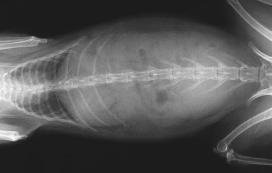
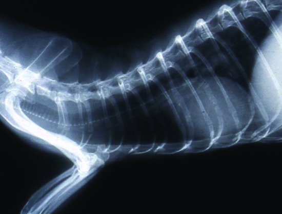
The caecum should always be full of ingesta, often with small volumes of gas in the healthy rabbit, and is positioned ventrally and to the right side. Excessive gas build-up such as that seen in mucoid enteropathy and penicillin toxicity where the whole of the stomach, small intestine, caecum and large intestine may be filled with fluid and gas (see Figure 7.3).
Figure 7.3 Gastric and intestinal bloat in a rabbit as a result of penicillin toxicity. Chest cavity is normal. The radiodense object over the neck is a staple in a skin wound. (Fraser and Girling, 2009)
The liver is found completely underneath the caudal ribcage and is a flattened shadow cranio-caudally. A number of liver diseases may result in enlargement and extension of the liver beyond the ribcage (e.g. hepatic lipidosis, hepatic coccidiosis due to Eimeria stiediae).
The stomach should always contain food – but a small gas crescent dorsally may be seen. Excessive gas build-up in the stomach can indicate a blockage or stasis of the bowel. Stomach emptying and intestinal motility are difficult to assess in the rabbit due to the permanently full stomach. However, one study suggested that 32% of a liquid marker should reach the caecum in 1 hour and 80% by 12 hours in the healthy rabbit (Pickard & Stevens, 1972). Solid markers however reached the caecum within 4 hours.
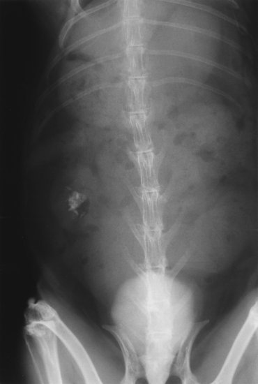
The normal kidney shadow should be 1.4–2.2 times the length of the second lumbar vertebra with a mean of 1.8 times. Renal calculi are frequently seen and may be a cause of intense pain in rabbits (see Figures 7.4 and 7.5). Negative contrast techniques after catheterisation of the bladder using air at 5–8 mL/kg body weight may be used in rabbits to outline the bladder lining. Double contrast techniques can be used with iodine at a rate of 2–3 mL/kg. Excretory urograms may be useful in assessing renal disease using 2 mL/kg of an intravenous iodine-based contrast media injected into a peripheral vein. In healthy rabbits, the presence of large amounts of calcium ‘silt’ may lead to a significant outline of the bladder.
Figure 7.4 Lateral abdomen of a rabbit showing radiodense calculi in left kidney and radiodense silt in bladder. Note also spondylosis lesions in L5–6 and L6–7. (Fraser and Girling, 2009)
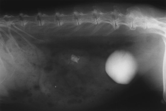
Figure 7.5 Dorsoventral view of the rabbit in Figure 7.4 showing multiple radiodense calculi in left kidney and radiodense silt in urinary bladder. (Fraser and Girling, 2009)
The uterus may become enlarged due to neoplasia (e.g. uterine adenocarcinomas), pyometra or gravidity. The vagina may also become enlarged due to venous aneurysms.
In the male rabbit there is no os penis. The testes sit scrotally but can be retracted into the abdomen.
Head
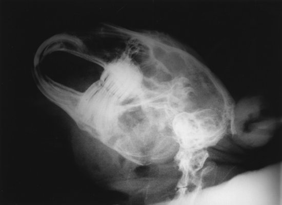
Radiography is essential to assess dental disease which is common in rabbits (Harrenstein, 1999) (see Figure 7.6). Root elongation and lysis of the alveolar bone is often an early radiographic sign of abnormal dental wear (Redrobe, 2001). Irregular wear of the teeth and touching of the cheek teeth at rest are also indicators of dental disease.
Figure 7.6 Lateral head of a rabbit showing cheek tooth and incisor malocclusion. Note contact of the occlusal surfaces of cheek teeth. Roots of cheek teeth projecting through ventral mandible and upper/lower incisors meeting end on. (Fraser and Girling, 2009)
Final stage of dental disease includes abscess formation and osteomyelitis. Oblique views of the head may allow examination of one dental arcade from the other.
Exophthalmos may be due to cheek–tooth abscess, retrobulbar neoplasia and cranial mediastinal masses such as thymic neoplasia; therefore, chest radiographs are also necessary.
Dacrocystitis may be associated with dental disease, often due to incisor root elongation or infection. Contrast studies of the nasolacrimal ducts may be performed using iodine-based contrast media injected into the ventral tear duct punctum and show the pinching of the duct around the roots of the incisor.
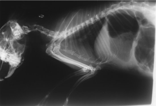
For otitis media causing vestibular disease, a dorsoventral square-on view of the skull is necessary (see Figure 7.7). Skull neoplasia has also been recorded in the rabbit (Weisbroth & Hurwitz, 1969).
Figure 7.7 Dorsoventral view showing middle ear disease in a rabbit (worse in right middle ear), dental disease (flaring laterally of first cheek teeth) and enlarged heart. (Fraser and Girling, 2009)
Appendicular skeleton
Osteoarthritis is common in rabbits. Luxation of the elbow has also been observed. Neoplasia of the limb in the form of fibrosarcomas and osteosarcomas are also seen in rabbits. Hypertrophic osteopathy has also been reported associated with an intrathoracic neoplasm (DeSanto, 1997), as has a limb neurofibrosarcoma in association with a thymic neoplasm (Clippinger et al., 1998).
Axial skeleton and ribs
The spine shows some breed variation in numbers of vertebrae with some individuals possessing 13 thoracic vertebrae (Kozma et al., 1974). Fractures of the spine, associated often with metabolic bone disease, or luxations/subluxations of the vertebral joints are common.
Arthritis of the spine (spondylosis) and spinous process fractures are also common. In one study, three types of spinal lesions were observed in the rabbit nucleus pulposus of the intervertebral discs undergoing chondroid metaplasia, eventually involving the whole spinal column by 2 years of age; mineralisation with hydroxyapatite deposition in the nucleus pulposus of the discs radiographically, primarily in the thoracic segments (seen as young as 3 months of age) and finally, spondylosis in rabbits over 24 months, chiefly bridging those discs that did not show mineralisation (Green et al., 1984).
Myelography has been described (Longley, 2005). The access point is L5/6 with 0.4 mL/kg of iodine-based contrast medium will extend the media from T2–L7. A 23G, 1¼-inch needle will allow access to the subarachnoid space.
Interpretation of rodent and marsupial radiographs
Imaging techniques in rodents and marsupials are the same as for other mammals. Their small size precludes the use of grids. The use of non-screen film may help in examining fine structures. In many cases, short exposure times are required due to the rapid rates of respiration, even when anaesthetised.
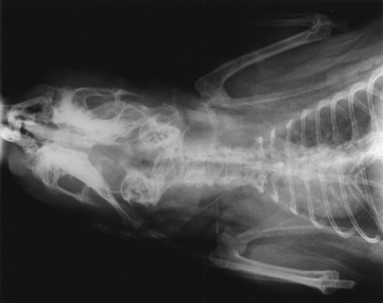
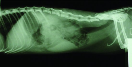
Anaesthesia or sedation is generally required in all cases to allow proper positioning. In all cases, several important features of rodents are visible on radiographs. The main problem is that the chest cavity is very small in relation to the abdominal cavity, making examination of the lung fields for minor abnormalities difficult (Girling, 2002) (see Figures 7.8a–7.8b). Generally though, when patients are presented, the pathology is advanced and it may be difficult to see any normal lung tissue! (see Figure 7.9). Heart disease is commonly seen in many small mammals (see Figure 7.10). Some species, such as rats, have open growth plates on many long bones, even as adults. Males of most rodents have an os penis, and extensive testicular tissue, which when retracted, may fill the caudal abdomen (see Figure 7.8a). In hind gut fermenters, such as the guinea pig and chinchilla, the capacious large intestine and caecum fills the abdomen making discernment of other structures difficult.
Figure 7.8 (a and b) Lateral and dorsoventral view of a normal healthy adult male rat. Note the small size of the chest, the open growth plates particularly on the proximal tibia and the ischial region of the pelvis and the presence of an os penis.
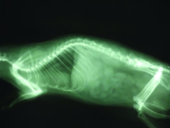
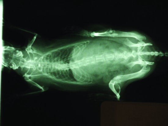
Figure 7.9 Lateral view of an adult (overweight) rat. Note the radiodense masses in the chest typical with mycoplasma pneumonia and lung consolidation. Note also the enlarged heart due to cor pulmonale.
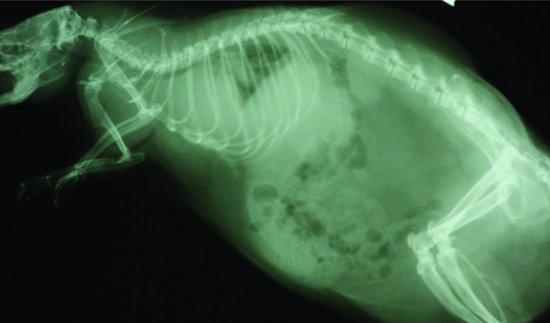
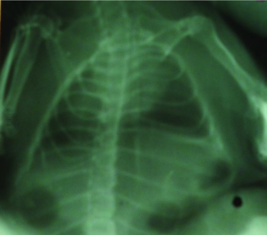
Figure 7.10 Dorsoventral view of a hamster’s chest with cardiomegaly due to heart failure associated with bacterial endocarditis. This radiograph was taken using dental non-screen film to improve minor details.
Dental disease is particularly common in Hystricomorphs such as chinchillas (see Figures 7.11 and 7.12), guinea pigs (see Figure 7.13) and degus.
Figure 7.11 Lateral view of a chinchilla. Note the capacious intestinal mass full of ingesta, the small lung field size, which makes the heart look enlarged. The dentition of this chinchilla is near normal.
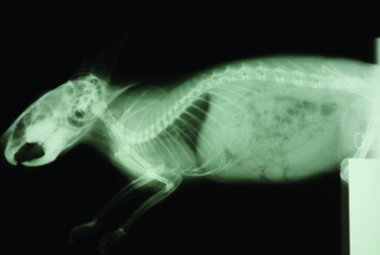
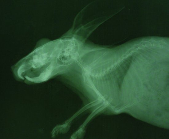
Figure 7.13 Lateral view of a guinea pig’s head and thorax. Note the small lung field size that makes the heart look enlarged. Note also in this case the elongated cheek teeth whose roots are pushing into the nasal passages and through the ventral mandible and the incisors, which are meeting end on end instead of the maxillary incisors meeting rostral to the mandibular incisors.
Marsupials have an epipubic bone projecting cranially from the pubis and thought to support the ventral body wall and pouch. The sugar glider however does not possess this.
Interpretation of ferret radiographs
Thoracic cavity
Heart
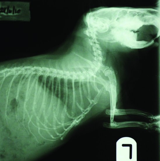
The heart is positioned more caudally than in the cat or dog and may be slightly elevated from the sternum due to fat deposition in the pericardiac ligament (Orcutt, 1998) (see Figures 7.14 and 7.15).
Figure 7.14 Lateral view of the thorax of a normal ferret. Note the apparently more caudal position of the heart and the narrow chest inlet which makes the trachea appear enlarged.
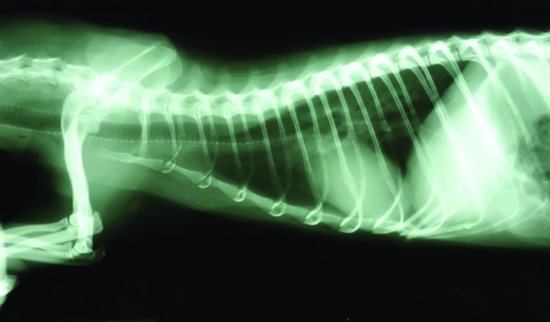
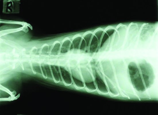
Figure 7.15 Dorsoventral view of the thorax of a normal ferret. Note the more transverse position of the heart. Note also in this case some gas in the stomach which in this case is due to pre-anaesthetic starvation.
Cardiac enlargement due to cardiomyopathy and congestive heart failure is common in ferrets (see Figure 7.16). The size of the heart may be assessed using the modified vertebral heart score (VHS) that compares heart length along the long axis against its width on a right lateral plain radiograph and relating this to the length of the heart in thoracic vertebral units similar to that seen in the dog and cat. The following formula has been used to assess the heart of both male and female ferrets:
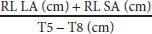
where RL LA means right lateral long axis (base of heart to apex), RL SA means right lateral short axis (widest part of the heart) and T5–T8 means thoracic vertebra 5 to thoracic vertebra 8.
The ratio was 1.35 (standard deviation 0.07) for males and a mean of 1.34 (standard deviation 0.06) for females (Stepien et al., 1999).
Heartworm infestation may show a pleural effusion and cardiomegaly, principally the right side of the heart. In addition, the caudal vena cava is often noticeably dilated (Supakorndej et al., 1995).
Lungs and other organs
Pneumonia produces a typically interstitial and then alveolar pattern similar to that seen in cats and dogs. Lung patterns around the perihilar region suggest lung oedema and congestive heart failure.
Pleural effusions may be seen due to a right-sided failure, for example thymic neoplasia, traumatic wounds and pleurisy or as with heartworm infestation. Soft tissue mineralisation due to over-supplementation with calcium and vitamin D3 can affect the primary vessels, and of course the kidneys.
The oesophagus
The oesophagus is not easily seen in the healthy ferret, but megaoesophagus has been reported. Contrast studies using barium sulphate are necessary to help define the oesophagus. A dose of 10–15 mL/kg orally and may be made more palatable by mixing it with a meat-based dog/cat food. Pollock (2007), however, describes that strawberry-flavoured barium sulphate is readily accepted by ferrets.
Abdominal cavity
Liver
The liver is normally completely covered by the caudal ribcage. Enlargement of the liver shadow has been associated with neoplasia (such as lymphoma, biliary cystadenoma, cholangiosarcomas and hepatocellular carcinomas), polycystic disease and infectious disease such as mycobacteriosis (Saunders & Thomsen, 2006).
Stomach and intestines
The stomach is on the left side dorsoventrally and immediately behind the dorsal part of the liver shadow on the lateral view. The cranial border of the stomach in a ventrodorsal radiograph extends to the 13th thoracic vertebra (Evans & An, 1998). Normally, the stomach has small amounts of gas present except in the case of an anaesthetic. Gastric bloat can be seen in recently weaned ferrets and can produce large amounts of gas (Fox, 1988). Other causes of gas in the stomach are usually due to ulceration of the stomach (e.g. foreign bodies, neoplasia and Helicobacter mustelae infections) or small intestinal foreign bodies.
The small intestine is much the same diameter throughout. There is no caecum.
Thickening of the small intestine may be seen due to neoplasia such as malignant lymphoma and inflammatory disease such as proliferative ileitis caused by the bacterium Lawsonia intracellulare. Alternatively, it may be more diffuse as in eosinophilic enteritis and inflammatory bowel disease.
Spleen
As mentioned, the normal spleen may appear larger in the ferret and so radiographic interpretation of disease in the spleen may be difficult. Gross splenic enlargement may occur due to neoplasia such as lymphoma (see Figure 7.17) or haemangiosarcomas.
Stay updated, free articles. Join our Telegram channel

Full access? Get Clinical Tree


