chapter 16 Small Animal Spine
To obtain a diagnostic radiograph of the vertebral column, two factors must be considered. First, the vertebral column must always be as parallel to the tabletop as possible. Second, the disk spaces of the spine must be nearly perpendicular to the tabletop and in parallel alignment with the central axis of the primary x-ray beam. These criteria can be met through a number of means.
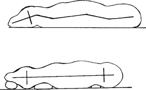
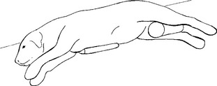
On rare occasions no manual assistance may be necessary to achieve correct positioning of animal patients placed in recumbency. However, it is usually necessary to alter the lateral recumbent position of the animal and positioning devices such as foam sponges, sandbags, orcotton may be helpful. Usually, efforts to improve the patient’s positioning focus on elevating the sternum and hind legs and providing support for the skull and midcervical and midlumbar regions (Figs. 16-1 and 16-2). Remember, any positioning device superimposed on an area of interest must be radiolucent.
Ventrodorsal View
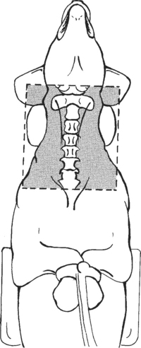
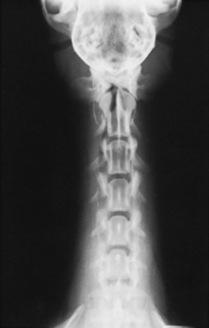
The patient is placed in dorsal recumbency with the head extended cranially and the front limbs pulled caudally alongside the body (Figs. 16-3 and 16-4). The patient must be restrained in a true ventrodorsal posture, and the cervical spine must be parallel with the cassette. A sponge pad or cotton can be placed under the midcervical region to eliminate any distortion in this area. The field of view should include the base of the skull, the entire cervical spine, and the first few thoracic vertebrae.
Extended Lateral View
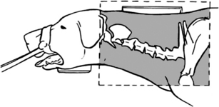
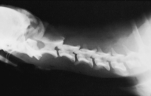
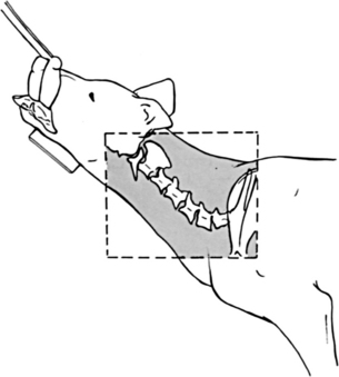
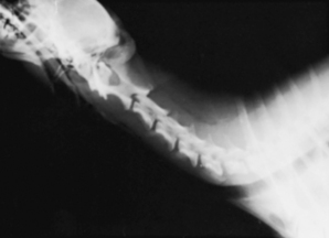
The patient is placed in lateral recumbency with the head and neck extended and the front limbs pulled in a caudal direction. Gentle traction should be placed on the cervical region by pulling the head of the patient in a cranial direction. This traction can be accomplished manually by stretching the cervical spine, or a length of roll gauze can be tied around the nose behind the canine teeth and pulled cranially (Figs. 16-5 and 16-6). A foam wedge pad is placed under the mandible to eliminate skull obliquity. To position the cervical spine parallel with the cassette, it may be necessary to place a sponge wedge pad or cotton under the midcervical region. The field of view should include the base of the skull, the entire cervical spine, and a few thoracic vertebrae.
BEAM CENTER: Over C4-5 intervertebral space
MEASUREMENT: C5-6 intervertebral space
BEAM CENTER: Intervertebral space of C-4 and C-5
Flexed Lateral View
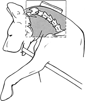
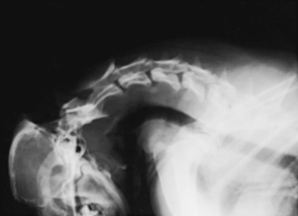
The patient is placed in lateral recumbency with the front limbs pulled in a caudal direction. A length of roll gauze or rope is tied around the mandible behind the canine teeth, and the free end of the line is placed between the forelimbs. Gentle traction is placed on the free end of the gauze, and the head is pulled caudally toward the humeri (Figs. 16-7 and 16-8). Care must be taken not to hyperflex the neck, which may cause tracheal trauma or collapse of an endotracheal tube. It may be necessary to elevate the vertebrae to the level of the thoracic spine with a sponge wedge pad or cotton. An appropriately sized cassette should be used so that the field of view includes the area from the base of the skull to the first few thoracic vertebrae.
BEAM CENTER: C3-4 intervertebral space
Hyperextended Lateral View
The patient is placed in lateral recumbency with the front limbs extended caudally. The head and neck region is extended in a dorsal direction until resistance is met (Figs. 16-9 and 16-10). A foam wedge pad or cotton is placed under the mandible to alleviate skull obliquity and under the midcervical region to align the vertebrae. The field of view should include the area from the base of the skull to the first few thoracic vertebrae.
BEAM CENTER: C3-4 intervertebral space
Stay updated, free articles. Join our Telegram channel

Full access? Get Clinical Tree


