chapter 17 Small Animal Soft Tissue
INTRODUCTION
The term soft tissue describes the areas of the body that surround the skeletal structures. Unlike radiography of bone tissue, visualization of soft tissue can be difficult because it involves only slight differences in radiographic density. Production of a soft tissue radiograph that has high contrast between the various adjacent soft structures is almost impossible without the use of contrast media. To achieve the correct contrast, density, and visualization, a number of factors must be considered:
Lateral View
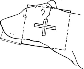
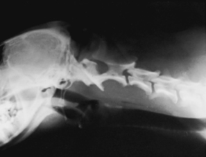
The patient is placed in lateral recumbency with the forelimbs pulled in a caudal direction. The head and neck are extended cranially and placed in a true lateral position (Figs. 17-1 and 17-2). A sponge wedge pad placed under the mandible helps eliminate obliquity of the skull and frees the larynx from the mandible to allow better visualization of the laryngeal region. The air passages of the upper respiratory tract act as a negative contrast agent and permit the soft tissue structures of the pharyngeal region to be differentiated. The field of view should include the entire area of the neck between the lateral canthus of the eye and the third cervical vertebral body.
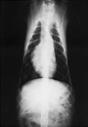
Dorsoventral View
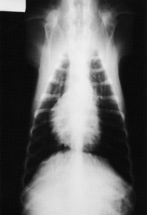
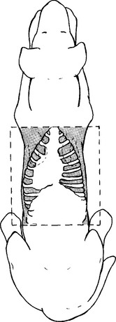
The patient is placed in sternal recumbency with the thoracic vertebrae superimposed over the sternum (Figs. 17-3 and 17-4). The forelegs are pulled slightly forward to prevent the elbows from tucking under the thorax. The rear legs are allowed to flex in a natural crouching position. This crouched position may be difficult for the canine patient with hip dysplasia, and it may be necessary to consider the ventrodorsal view. The head is lower and is placed between the two forelimbs. The field of view should include the entire thorax. The rule is “the thorax is inside the rib cage”; if you include all of the ribs, you will radiograph the entire thorax.
BEAM CENTER: Over caudal border of scapula
Ventrodorsal View
The ventrodorsal view of the thorax is advocated when a full view of the lung fields is necessary. This projection provides a better view of the accessory lung lobes and caudal mediastinum. Although it is easier to control a patient in ventrodorsal position, this view is contraindicated for patients in obvious respiratory distress. Placing such an animal on its back would be dangerous and could possibly cause further respiratory problems.
The patient is placed in dorsal recumbency with the forelimbs extended cranially (Figs. 17-5 and 17-6). The hind limbs can assume a normal position. Great care must be taken to ensure that the patient is in a true ventrodorsal posture. The sternum must be superimposed over the spine. If rotation is encountered, a V trough or sandbags placed under the pelvic region may be helpful. The field of view should include the entire thorax. The rule is “the thorax is inside the rib cage”; if you include all of the ribs, you will radiograph the entire thorax.
BEAM CENTER: Over caudal border of scapula

