chapter 2 Anatomy of the X-ray Machine
Upon completion of this chapter, the reader should be able to do the following:
Acceleration: The increase in speed over time.
Actual focal spot: The area of the focal spot consisting of a coiled wire that is perpendicular to the surface of the target.
Anode: A positively charged electrode that acts as a target for the electrons from the cathode. Electrons interacting with the anode produce heat and x-rays.
Arcing: A phenomenon in which metal deposits on the inner wall of the envelope act as a secondary anode, thereby attracting electrons from the cathode.
Autotransformer: Provides a variable yet predetermined voltage to the high-voltage step-up transformer. It acts as the kilovoltage selector.
Cathode: A negatively charged electrode that provides a source of electrons.
Collimator: A restricting device used to control the size of the primary x-ray beam.
Console: The control panel of the x-ray machine.
Effective focal spot: The area of the focal spot that is visible through the x-ray tube window and directed toward the x-ray film.
Filament: Part of a low-energy circuit in the cathode that, when heated, releases electrons from their orbits.
Focal spot: The small area of the target with which electrons collide on the anode.
Focusing cup: A recessed area where the filament lies, directing the electrons toward the anode.
Full-wave rectification: Creates an almost constant electrical potential across the x-ray tube, converting the positive electrical current pulses to 120 times per second compared with the normal rate of 60 times per second.
Glass envelope: A glass vacuum tube that contains the anode and cathode of the x-ray tube.
Half-wave rectification: A method of converting alternating to direct current in which half of the current is lost.
Heel effect: A decrease of x-ray intensity on the anode side of the x-ray beam caused by the anode target angle.
Kilovoltage: The amount of electrical energy being applied to the anode and cathode to accelerate the electrons from the cathode to the anode (1 kilovolt [kV] = 1000 volts [V]).
Kilovoltage peak (kVp): The peak energy of the x-rays, which determines the quality (penetrating power) of the x-ray beam.
Line-focus principle: The effect of making the actual focal spot size appear smaller when viewed from the position of the film because of the angle of the target to the electron stream.
Line-voltage compensator: Adjusts the incoming line voltage to the autotransformer so that the voltage remains constant.
Milliamperage (mA): The amount of electrical energy being applied to the filament. Milliamperage describes the number of x-rays produced during the exposure.
Molybdenum: A metal commonly used in focusing cups because of its high melting point and poor conduction of heat.
Penumbra: Partial outer shadow of an object being imaged by illumination.
Rectification: Process of changing alternating current to direct current.
Rotating anode: An anode that turns on an axis to increase x-ray production while dissipating heat.
Stationary anode: A nonmoving anode, usually found in dental and small portable radiography units.
Step-down transformer: Reduces the x-ray machine input voltage from 110 or 220V to 10V to prevent burnout of the cathode filament.
Step-up transformer: Increases the incoming voltage of 110 or 220V to thousands of volts (i.e., kilovolts).
Timer switch: Controls the length of exposure.
Tungsten: A common metal used in the filament of a cathode.
Valve tubes: Allow the flow of electrons in one direction only. Commonly called self-rectifiers.
X-ray tube: A mechanism consisting of an anode and a cathode in a vacuum that produces a controlled x-ray beam.
THE X-RAY TUBE
X-ray Production
The following elements are necessary for x-ray production:
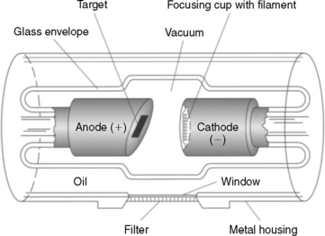
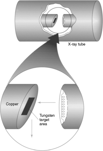
The x-ray tube consists of a cathode side (with a negative electrical charge) and an anode side (with a positive electrical charge) encased in a glass envelope, which is evacuated to form a vacuum (Fig. 2-1).
Cathode
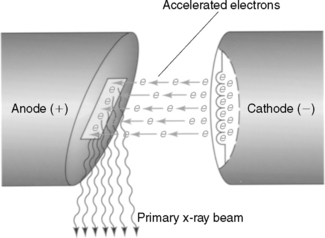
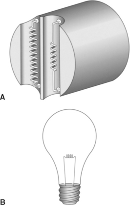
The purpose of the cathode is to provide a source of electrons and direct these electrons toward the anode (Fig. 2-2). The cathode consists of a coiled wire filament that emits electrons when heated. The filament in most x-ray tubes measures approximately 0.2 cm in diameter and 1 cm in length. It is mounted on rigid wires that support it and carry the electrical current that is used to heat the filament. The filament of the cathode is similar to the filament of a light bulb (Fig. 2-3). When a filament is heated, electrons are held less tightly by the nucleus of the atoms of the metal. In other words, the electrons become excited. When the energy level exceeds the binding energy, a cloud of electrons is formed and made available to travel to the anode.
The filament is constructed of tungsten because of its high melting point (3370°C) and high atomic number. The atomic number is the number of protons in the nucleus of an atom. This number is matched by an equal number of electrons traveling around the nucleus. A high atomic number is proportionate to the potential electron availability. A metal of this type is also necessary because of the great amount of heat produced at the filament. Some x-ray tubes, usually those used in small portable and mobile units, have a single filament. Most modern tubes have two filaments mounted side by side. One is smaller than the other, and each has a different capacity for heat and electron emission.
The filament is heated by a low-energy circuit. The amount of energy in the circuit is referred to as milliamperage (mA). As the milliamperage is applied and the filament is heated, electrons are released from their atomic orbits. The quantity of electrons produced depends on the heat of the filament. Because of its negative electrical charge, the electron cloud is attracted to the anode side of the tube. The electron stream must be accelerated to create an impact great enough to produce x-rays. Acceleration of the electrons is controlled by the kilovoltage applied between the anode and the cathode. Milliamperage and kilovoltage are discussed in more detail in Chapter 4.
Anode
Types of Anodes.
STATIONARY ANODE.
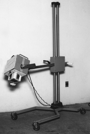
Stationary, or “fixed,” anodes are found in dental and small portable radiography units. These units have a relatively small capacity for x-ray production (Fig. 2-4). As shown in Figure 2-5, the tungsten target area of the stationary anode is embedded on a cylinder of copper, with the face of the target angled down toward the window. The angle may range from 15 to 23 degrees, altering the “focal spot” size. The focal spot is the small area of the target with which the electrons collide. The focal spot is discussed in detail later.
The primary limitation of the stationary anode is its inability to withstand large amounts of heat. Repeated bombardment by electrons and subsequent heat production can damage the target. Damage commonly seen from this repeated bombardment is a pitting of the target surface. Once a target has been damaged in such a way, the x-rays produced from that area scatter in undesirable directions (Fig. 2-6). Radiographs produced by an x-ray tube with a pitted target area appear lighter than expected.
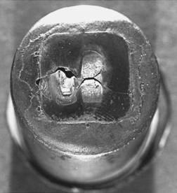
Figure 2-6 Pitted anode target area showing scatter radiation resulting from the uneven target surface.
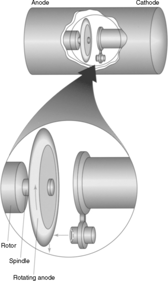
ROTATING ANODE.
The rotating anode is disk shaped and rotates on an axis through the center of the tube (Fig. 2-7). The disk is approximately 3 inches in diameter with a beveled edge. It is composed of tungsten or some similar alloy that can withstand high temperatures. The spindle on which the anode is mounted usually is made of molybdenum. Molybdenum dissipates the heat produced on electron impact. This heat reduction is necessary to reduce the heat flow to the rotor and bearing mechanism that spins the anode.
Stay updated, free articles. Join our Telegram channel

Full access? Get Clinical Tree


