CHAPTER 7 Skin Infections
Skin infections, especially contagious dermatologic diseases, include some of the most common causes of equine skin disease. In one report, 25% of all dermatologic diagnoses made over a 21-year period were infectious in origin.1 For the clinician, it is important to diagnose these conditions quickly to prevent spread to other horses and potentially their human companions. Many of these infectious agents are discussed in detail elsewhere in this book. The purpose of this chapter is to provide an overview that assists the clinician in differentiating, diagnosing, and treating conditions given the appropriate historical information, clinical presentation, and laboratory data.
GENERAL COMMENTS
A detailed history is an extremely important component of any dermatologic workup (Box 7-1). The age at time of onset, time of the year, presence of itching, and any other notable detail not generally observed during a short examination can assist physical diagnosis. Additional questioning should include whether any topical medications, home remedies, or systemic medications have been used. Response to medications (independent of season) and time to relapse (if a response occurred) are valuable pieces of information to assist diagnosis.
Dermatologists have a basic and straightforward diagnostic armamentarium that differentiates most diseases. Skin scrapings are essential for diagnosis of parasitic infections (see Chapter 64). Punch biopsies of skin are quick and inexpensive and can yield much needed information. Collection of a sterile biopsy for culture is necessary for bacterial and some fungal infections. When disease is global and long-standing, a wedge biopsy may yield more information regarding the primary etiology. Accurate histopathologic assessment with appropriate knowledge of equine-specific diseases is essential. For example, horses develop sarcoids frequently and fibrosarcomas rarely, and the two conditions are treated very differently (see Chapter 25). Special stains for bacteria and fungi should be pursued when appropriate.
CRUSTING/SCALING DERMATOSES
Bacterial Etiologies
Crusting/scaling dermatoses affect primarily the epidermis and upper levels of the dermis and have many causes. Often these dermatoses start as small papules that quickly progress to crust and scale. In addition, clinical signs may include small papules, erosions, excoriations, alopecia, and varying degrees of pruritus (Boxes 7-2 and 7-3).
Bacterial folliculitis is most often caused by staphylococcal bacteria (Staphylococcus aureus, S. intermedius, S. hyicus).2,3 Other bacteria infrequently associated with folliculitis include Streptococcus equi subsp. equi, Streptococcus equisimilis, and Corynebacterium pseudotuberculosis.4–6 Dermatophilosis is another bacterial crusting/scaling dermatosis in the horse (see Chapter 31).
Clinical Findings
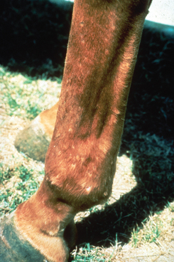
Most cases of folliculitis start in the spring or summer. The lesions start as small follicular papules that can develop into transient pustules that rupture to form crusted pustules (Fig. 7-1). Small foci of alopecia with scaling develop in areas of infected follicles, leading to a moth-eaten appearance of the hair coat. In severe cases, lesions may enlarge, ulcerate, and have a purulent or serosanguineous discharge. If complicated by furunculosis, nodules and fistulous tracts may be present. Papular lesions can be more painful than pruritic. Most of these lesions start in the tack areas and are associated with friction, high temperature and humidity, excessive sweating, poor grooming practices, and possibly biting insects. Horses also develop staphylococcal folliculitis of the caudal aspect of the pastern and fetlock, which is considered to be one of the causes of “grease heel.” If left untreated, lesions of folliculitis may become widespread.
Therapy
Treatment consists of systemic antibiotics, topical therapy, and improved grooming practices and attempts to eliminate other predisposing factors. Antibiotic selection should be based on results of culture and sensitivity testing. This is especially true now that methicillin-resistant Staphylococcus aureus (MRSA) infections that potentially can be transferred to horse personnel are being diagnosed more frequently in horses7–9 (see Chapter 29). Antibiotic therapy should last at least 3 to 4 weeks, or 7 to 10 days beyond clinical cure. Topical therapy may consist of chlorhexidine or benzoyl peroxide shampoos. Benzoyl peroxide has the most antibacterial activity but may dry and bleach the coat. Therefore the frequency of topical application depends on the severity of disease and the topical agent used. For most cases, once-weekly to every-other-week application is adequate.
Prevention
Improved hygiene is required both for prompt resolution of infection and for prevention of recurrences. All blankets should be thoroughly cleaned with hot water and bleach. Tack should be kept as clean as possible. Cleaning of equipment should be continued on a routine basis. The horse should not be ridden while lesions are active. After the lesions have improved, the horse should be cooled properly and rinsed well after each ride. The horse should be bathed once or twice weekly in hot weather and should always have a clean saddle blanket each time it is ridden.
Fungal Etiologies
Dermatophytosis is a common cause of crusting and scaling (see Chapter 54). Malassezia has recently been associated with crusting/scaling dermatitis in the horse.10 Malassezia pachydermatis is a lipophilic, nonmycelial, saprophytic yeast often found on normal and abnormal skin of various mammals, including the horse. A novel Malassezia species also inhabits normal horse skin. This organism has not been completely characterized but is closely related to Malassezia sympodialis. As in the dog and cat, Malassezia in the horse tends to be associated with other dermatologic diseases, including atopy, insect bite allergy, food allergy, and dermatophytosis.


