CHAPTER 54 Dermatophytosis
Dermatophytosis (“ringworm”) is a common superficial cutaneous fungal infection caused by keratinophilic fungi that are able to invade the stratum corneum of the skin and other keratinized structures. Several dermatophytes are reported to induce cutaneous disease in horses. Clinically, dermatophytosis presents as a crusting and scaling disease, similar to bacterial folliculitis.
ETIOLOGY
Trichophyton equinum is the most common causative agent of dermatophytosis of horses.1,2 Two varieties of T. equinum have been reported, T. equinum var. equinum and T. equinum var. autotrophicum. Other, less common agents are Trichophyton verrucosum, T. mentagrophytes, Microsporum equinum, M. canis, and M. gypseum.3–5 In one study, T. equinum var. autotrophicum, M. canis, and M. equinum were reported to be restricted to racing horses only, whereas M. gypseum occurred in racing, riding, and breeding horses.6
EPIDEMIOLOGY
Dermatophytes are highly contagious. Transmission occurs by either direct contact between horses or through contact with contaminated equipment. Infection can be readily transmitted, particularly by infected saddle-girths, on which the fungus can survive for 12 months.7 Insects are also reported to play a role in the transmission of disease.8,9 The source of infection varies depending on the dermatophyte. Some dermatophytes are zoophilic, and the source of infection may be another infected animal, such as a horse (e.g., T. equinum), a cat (e.g., M. canis), cattle (e.g., T. verrucosum), or a rodent (e.g., T. mentagrophytes). Other dermatophytes are geophilic (e.g., M. gypseum), and the source of infection is infected soil.
The prevalence of ringworm varies greatly depending on geographic location and husbandry conditions. In one epidemiologic study conducted in Egypt, 42% of horses with skin disease were positive for dermatophytes.10 Horses less than 2 years old were more susceptible to infection. Fourteen species belonging to nine genera of keratinophilic and cycloheximide-resistant fungi were recovered from collected specimens. Trichophyton was the dominant genus, and T. equinum was the most frequently identified dermatophyte.
In a study of 200 horses in Italy, only 9% of horses were positive for T. equinum.11 Although the clinical diagnosis of “ringworm” is quite common, many cases that clinically resemble ringworm may be bacterial folliculitis. At the University of Florida, only 5% of all the horses with skin disease that have cultures for dermatophytes are confirmed to have ringworm. The majority of horses that clinically appear to have ringworm are instead diagnosed with bacterial infections, either Staphylococcus or Dermatophilus.
PATHOGENESIS
Virulence Factors
Dermatophytes produce enzymes such as keratinases that enable invasion of the hair and the stratum corneum and facilitate establishment of infection.12,13 T. equinum produces urease, gelatinase, protease, hemolysins, and keratinase.14 Some differences in enzyme production have been found between T. equinum and T. mentagrophytes. T. mentagrophytes may have stronger enzymatic properties, which, clinically, lead to more inflammatory reactions. Hemolytic activity and the ability to induce hypersensitivity reactions are also important virulence factors, especially for Trichophyton species.15,16
Host Factors and Immune Response
The conditions of the skin and immune system of the host play a crucial role in determining whether infection is established and how readily it is eliminated. Any impairment of the barrier function of the skin may foster establishment of infection. In particular, abrasions can facilitate the development of lesions and prolong the recovery period; abrasions are an important risk factor for infections in the girth area.6,7
Young horses (<3 years of age) are at increased risk for developing dermatophytosis.6,7 Stress, such as training, also predisposes to the development of ringworm. In one study, 32% of horses in training were clinically affected, whereas only 1.1% of breeding horses were affected with pathogenic dermatophytes.6 Concurrent diseases that may compromise the immune response can predispose to the development of dermatophytosis.17
Immunity is acquired by active infection. Both nonspecific and specific immune responses are important for clearing dermatophyte infections. Serum inhibitory factors deprive dermatophytes of iron, which is an essential nutrient. Both humoral and cell-mediated immune responses are elicited, but cell-mediated immunity appears to be most important for resolution of infection.18,19 The development of cell-mediated immunity correlates with the development of an inflammatory response and is associated with clinical cure, whereas the lack of or a defective cell-mediated immunity predisposes the host to chronic or recurrent dermatophyte infection.20 The inflammatory reaction also promotes keratinocyte proliferation, which facilitates the elimination of the fungus from the skin surface.
Horses that self-clear dermatophyte infections develop immunity and rarely experience recurrence of infection. Some horses, because of either a defective immune response or a concurrent illness, fail to develop long-term immunity and are prone to recurrent infection, unless the dermatophyte is completely eliminated from their environment.
CLINICAL FINDINGS
Primary lesions of dermatophytosis consist of follicular papules and pustules. Individual lesions may present as spreading circular patches of alopecia (Figs. 54-1 and 54-2), surrounded by erythema and scaling (epidermal collarettes). Urticaria-like lesions can be observed in early stages of the disease. As the infection progresses, crusting and scaling (seborrhea) develop. Pruritus is usually absent but may be present in some cases. Hair is epilated easily in affected areas. In some horses, nodular lesions can develop as the result of ruptured follicles (furunculosis), which elicits a strong inflammatory response, intense erythema, and suppurative exudate.
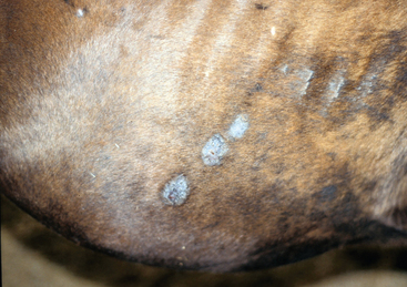
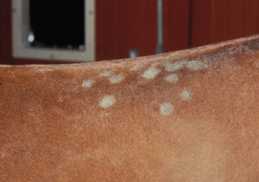
The most frequently affected sites are the girth and the shoulder area, usually from use of contaminated equipment. A survey of 568 horses in training and 2535 horses on breeding farms showed that the majority of lesions on racing horses were located on the girth areas.6 Other frequently affected areas are the muzzle and the pastern region.
Stay updated, free articles. Join our Telegram channel

Full access? Get Clinical Tree


