Skin Diseases
7.1 Underlying causes of skin disease in rabbits
Skin disease is common in pet rabbits but apparently rare in their wild counterparts. The wild rabbit has a short dense coat that it grooms regularly using the incisors to pull out dead hair. The coat is short and dense and does not knot and mat easily like the fluffy coats of many pet rabbits. Wild rabbits live in groups where mutual grooming is an important part of their social behaviour. During periods of rest, they lie together and groom each other, especially around the face and head. Wild rabbits are not confined to a small space and do not sit for hours on end on a bed contaminated by urine or faeces. They do not become obese and disabled individuals are quickly caught by predators.
Pet rabbits who may live alone and suffer any or all of these problems rapidly show signs of skin disease due to lack of ability to groom. Solitary rabbits have no companion to groom them. The fast-growing, fine fluffy coat of breeds such as the angora is impossible for a rabbit to groom. Incisor malocclusion removes the pincer-like action of the teeth, making removal of dead hair difficult. Cheek tooth malocclusion results in the formation of sharp spurs that grow into the tongue, making licking and grooming painful. Effective grooming requires flexibility, which is impaired by obesity, arthritis or spondylosis. Kyphosis, scoliosis and spondylosis are often found in pet rabbits (see Section 10.7). Underlying neurological problems, such as encephalitozoonosis, affect the ability to balance and adopt the correct posture to reach inaccessible parts of the body. Rhinitis makes it hard to breathe and groom at the same time. Hard flooring, inactivity, obesity and poor conformation predispose to avascular necrosis of the skin over the bony prominences of the feet and the development of sore hocks. Abrasive surfaces such as wire cages or tough carpets can traumatize the skin and predispose to infection. Damp, dirty bedding encourages bacterial contamination of skin and the array of chemicals, such as disinfectants and shampoos, that pet rabbits are exposed to increases the incidence of contact dermatitis and allergies. Some present-day breeds, such as the French lop, develop huge skin folds under the chin or around the genitalia, which are prone to moist dermatitis.
The key to successfully treating rabbit skin disease is to identify and, if possible, treat the underlying reasons, in addition to treating the condition itself. Many skin diseases can be alleviated or prevented by the provision of a soft clean bed, the opportunity to exercise, a high fibre diet and a companion, maybe human, who will diligently groom the coat, keeping it free from mats and debris.
7.2 Examination of the skin
The approach to skin disease follows the same principles as in other species. Anamnesis should include diet and husbandry regimens and in-contact animals. A full clinical examination, including the mouth, perineum and hocks may reveal underlying conditions that interfere with normal grooming activity and result in skin disease. The skin of mature entire male rabbits can become thickened along the dorsum from the neck to the rump, making injections difficult to impossible in this area. Histologically, the skin shows prominent, thick, dermal collagen similar to the cheek skin from entire male cats (Hargreaves and Hartley, 2000; Mackay, 2000). This is a secondary sexual characteristic.
The conformation of the rabbit is important. Some rabbits develop large skin folds under the chin or around the perineum, which can become infected. Large, loose eyelids (as seen in French lops) can interfere with the protective mechanism of the precorneal tear film and result in epiphora and facial dermatitis. Some rabbits develop a large ‘skirt’ of skin around the thighs that is in constant contact with the ground and prone to mechanical trauma and contact dermatitis. Poor conformation or mobility problems due to spondylosis or obesity can lead to pressure sores or the inability to adopt the correct position for urination, which results in urine scalding (see Figure 12.1).
The rabbit needs to adopt a variety of postures to reach all areas of the body. The face and ears are groomed using the front legs (Figure 7.1). The area between the tail and the dorsum and the area between the shoulder blades at the back of the neck are the most difficult parts of the body for a rabbit to reach (see Figure 1.19). A bonded companion will groom its mate, especially around the head and face but seldom around the tail (Figure 7.2). Combing through the coat with a fine-toothed flea comb gives an idea of whether the rabbit is grooming effectively. If dead hair can be groomed or pulled out of all areas of the body, then it can be assumed that the rabbit is hardly grooming at all and is suffering from a generalized condition. If some areas are well groomed and the ‘difficult to reach’ areas are not, then a flexibility problem may be present. The amount of dead hair increases during periods of moulting.
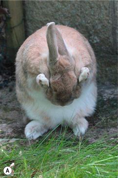
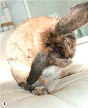
Figure 7.1 (A and B) Normal grooming positions.
It is clear from these photographs that normal grooming requires a rabbit to be mobile and flexible and have good balance. As soon as the mobility is affected, due to either pain or injury, or the balance is affected due to middle ear disease or central nervous disease, a rabbit’s ability to groom normally is compromised. This results in an unkempt coat; a proliferation of mites may be noted and urine scalding or failure to eat caecotrophs may also be seen. As well as addressing the obvious skin issues, the underlying causes should be sought and treated appropriately. Images courtesy of Heather Pinchien.
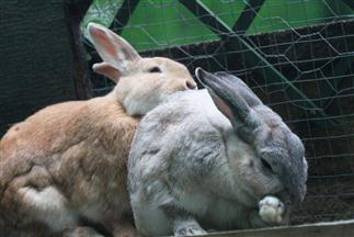
Figure 7.2 A bonded pair of rabbits grooming.
The rabbit at the front is grooming her face, while her companion grooms her back.
Good illumination and magnification facilitate close examination of the fur. Broken hairs, flea dirt or mites may be seen, especially in white fur. Infestation with Leporacarus gibbus (fur mite) gives the impression that the coat has been dusted with ‘salt and pepper’. Evidence of trauma, alopecia and erythema can all be seen on close examination of the skin. Some rabbits start to lick and chew at the area under the neck or may lick the handler in response to being combed, especially along the dorsum at the base of the tail. This response can indicate an underlying pruritic condition in an inaccessible site. For example, obese rabbits or those that cannot reach their perineum due to spondylosis may start to lick their dewlap, in response to being combed, especially if they are affected by perineal dermatitis or cheyletiellosis at the base of the tail.
There are many laboratory aids to diagnosis of skin conditions in rabbits (see Section 2.7).
7.3 Grooming and dematting rabbits
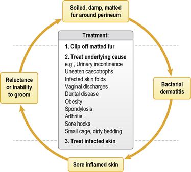
Grooming is important in rabbits to keep the coat free from debris and dead hair, and to prevent the formation of mats. Fluffy and long-haired breeds are especially prone to developing mats between the hind legs and around the base of the tail. Dirty bedding, or urine or faecal contamination, can result in the skin beneath these mats becoming infected and inflamed. Clipping away the soiled, matted hair and treating the underlying skin breaks a vicious circle that occurs when the coat is too matted and the skin too painful for the rabbit to groom properly, but lack of grooming allows mats to form and become soiled (see Figure 7.3). Some mats can be teased apart and the dead hair gently pulled out before combing through the remaining fur. It is important to be gentle when pulling mats out of the fur as the skin can be torn easily. Solid mats need to be cut out. A sharp pair of curved scissors can be used, either to cut through the mat perpendicular to the skin in several places, allowing it to be brushed out more easily, or to remove the entire mat, taking care to cut close to the mat without cutting the skin. Time, patience and good illumination are required. Many rabbits require sedation and pain relief (see Box 4.4). Clippers can be used to remove matted fur, but are often unsatisfactory for the removal of consolidated mats. The fine hair quickly becomes trapped in the blades. Stretching the skin out in front of the clippers and running them slowly over the skin reduces the risk of traumatizing the skin.
Bathing soiled rabbits is seldom satisfactory without clipping away the soiled fur first. It is almost impossible to cleanse the dense, fine fur of a rabbit, which usually becomes matted as a result of bathing. A mass of soiled, damp fur overlying infected skin is counterproductive. Bathing rabbits can be stressful and there are reports of shock and death of rabbits following the use of insecticidal baths or dips (Harvey, 1995). Clipping and cleansing affected areas is often effective without the use of baths. Hair needs to be removed as it is a barrier between the skin and any topical preparations that are applied.
7.4 Moulting
Rabbit hairs arise singly or in multiples from the hair follicles (Sandford, 1996), giving the characteristic dense coat. Hair grows quickly on newborn rabbits and initially consists of guard hairs followed by the soft undercoat over a matter of a few days. The baby coat is replaced at 5–6 weeks by the intermediate coat, which persists until the rabbit is 4–5 months old. This intermediate pelt is free from moulting hairs and is used in the fur industry.
Moulting in adult rabbits follows a seasonal pattern and there are usually two complete coat changes per year. During moulting, there are areas of fur in various stages of growth throughout the body. The moult usually starts on the head and works down the neck and back, with the stomach being the last area to shed the coat. Environmental stimuli, hormones and nutrition affect the moulting process. The density of the coat is also affected by environmental temperature and nutrition. The summer coat is generally shorter than the winter coat and may vary in colour. Colour point breeds such as Californian or Siamese sable show a similar pigment response to exposure as Siamese cats, where hair regrowth in shaved areas may be coloured black (Cheeke et al., 1982).
7.5 Alopecia
Hair loss in rabbits can be physiological. Some rabbits are naturally thin coated in the area at the nape of the neck. The modified coat texture of breeds such as the angora, dwarf and miniature lop has resulted in fluffy, fine fur that knots and mats easily. It is often shed in patches rather than in the typical pattern that occurs in wild rabbits or coarser coated breeds such as the Dutch or English. Alopecic areas that can cause concern appear (see Figure 1.9), but regrowth is usually rapid with areas of dense new hair appearing within 7–10 days. This phenomenon of patchy hair growth appears to be a variant of normal moulting. It may be more apparent after an area has been clipped for surgery (Hoyt, 1998).
In pregnant does the hair loosens and is plucked to line the nest during late pregnancy. This behaviour can result in large bald areas appearing on the chest and ventral abdomen. Pseudopregnant does also pluck hair from these areas. A bonded companion can chew the fur of its cage mate to stubble, especially on the lateral flanks. This process is known as barbering. This condition is relatively uncommon, and while it may be caused by a dominant companion, Jenkins (2001) found it was often performed by a subordinate animal. Microscopic examination of the fur shows broken hair shafts.
Poor nutrition can underlie alopecia. Sulphur-containing amino acids are required for keratin production and wool growth. Rabbits have an absolute requirement for essential amino acids despite amino acid synthesis in the caecum (Cheeke, 1987): see Box 1.3. Keratin is characterized by its high content of cystine, which is synthesized from the essential amino acid methionine (McDonald et al., 1996). Lysine is also important in the formation of keratin as well as fibrin and collagen. In general, cereals are deficient in lysine and methionine, whereas legumes are good sources. Sulphur amino acid deficiency can be reflected in poor coat quality in rabbits that are debilitated or are fed on a high cereal diet. Selective feeding from mixed rations results in a diet consisting almost entirely of cereals. Magnesium deficiency has also been linked with alopecia and alterations in fur texture in rabbits (Cheeke, 1987).
Areas that have been traumatized, e.g., by injection reactions or fight wounds, can remain hairless for some weeks after the lesion has healed. When rabbit skin is healing there can be a noticeable variation in the rate of hair regrowth, with those areas having the greatest blood supply growing hair more rapidly. Repetitive trauma and contact dermatitis can result in patches of alopecia in areas of skin in contact with flooring, especially the hocks. If a contact reaction is present, the skin is thickened, inflamed, hyperaemic and pruritic.
7.6 Injection reactions
Rabbits are prone to injection reactions. A lesion is found at the site of the injection a few days later. In some individuals, the reaction is extensive and areas of epidermis can slough. Adjuvanted viral haemorrhagic disease vaccine, carprofen and enrofloxacin injections are common culprits. Injection reactions can be avoided by ensuring that the product is delivered subcutaneously and not intradermally and dispersed by gently massaging the skin. Although injection reactions are unsightly, most heal quickly. If necessary, topical antiseptic creams can be applied.
7.7 Dermatitis and bacterial skin disease
Rabbit skin is thin and easily traumatized. It is reactive and easily irritated. Primary irritant contact dermatitis can result from the application of chemical agents such as topical iodine washes (Wilkinson and Harvey, 1994). Superficial pyoderma is common and is often secondary to underlying causes such as epiphora, ptyalism, urine scalding, bite wounds, injection reactions and infected skin folds. Deep skin folds accumulate debris and provide the correct environmental conditions (warmth and moisture) for bacteria and yeasts to grow. Some rabbits have excessively large folds of skin around the anus and genitalia.
Staphylococcus aureus is a common pathogen in infected skin conditions in rabbits (DeLong and Manning, 1994). Staphylococcus aureus can be isolated from all body sites in rabbits, but is present in greater numbers in the ear and perineum. There are several different biotypes and phage types that are related to virulence (Hermans et al., 1999).
Other bacteria such as Fusobacterium necrophorum or Pseudomonas aeruginosa are also associated with skin infections, especially in farmed rabbits under intensive conditions. ‘Schmorls disease’ or necrobacillosis is caused by F. necrophorum and is characterized by necrosis, ulceration and abscessation. Fusobacterium necrophorum is commonly found in rabbit faeces and this disease results from faecal contamination of wounds. In severe cases underlying structures such as bone may become involved. Diagnosis should be based on bacterial culture, and treatment should comprise surgical debridement as well as systemic antibiosis.
Pseudomonas aeruginosa is associated with moist dermatitis of the dewlap in rabbits kept under damp conditions. The infection is characterized by a blue/green discoloration of the fur (‘blue fur disease’) and is associated with intensive husbandry, wet bedding and contaminated water bottles (Scott et al., 1995). This type of infection is rare in the pet rabbit; however, where there is severe dental disease and excessive salivation or perhaps faulty drinking bottles causing excessive wetting of the skin it may be seen. Diagnosis should be made based on clinical signs and cytology of skin touch preparations. Clipping, cleaning and occasionally systemic antibiosis will be required, although this should be based on culture and sensitivity. Identification and correction of underlying factors, for example dental disease, should also be undertaken.
Pasteurella multocida is the most common pathogen in purulent skin conditions. The organism can be found in the nares of healthy rabbits and can be spread to other sites during grooming. Pasteurella multocida is often cultured from abscesses (see Section 6.1) and is a cause of respiratory tract infections (see Section 11.2.1).
Superficial pyoderma is treated by clipping hair away from the lesion, cleaning the skin and applying an antiseptic or antibiotic cream. Bathing may be necessary to remove any exudate or faecal contamination but is counterproductive unless the overlying fur is clipped off. The dense coat of rabbits mats easily and dries slowly, and moist conditions encourage bacterial growth. If bathing is necessary, chlorhexidine or povidone/iodine preparations are suitable skin cleansers and are effective against yeasts that inhabit skin folds. It should be noted that iodine preparations can cause skin irritation. Treatment with topical preparations containing antibiotic and corticosteroid are effective. However, topical preparations containing steroids should be used with caution. Corticosteroids are absorbed from these preparations, especially if the skin is inflamed (see Section 3.10). Prolonged administration of topical corticosteroids can result in thinning of the skin, and systemic absorption can result in leucocyte lysis and impairment of immune function, allowing manifestation of previously subclinical disease (for example, pasteurellosis or ‘snuffles’). The use of topical steroids can be counterproductive in some cases. Products that contain fusidic acid are very useful and only need to be applied once daily. Fusidic acid does not interfere with the gut flora and is effective against the staphylococci species often present under infected skin conditions. Fuciderm (Leo Laboratories), which contains steroids, and Fucidin (Leo Laboratories), which does not, are both suitable.
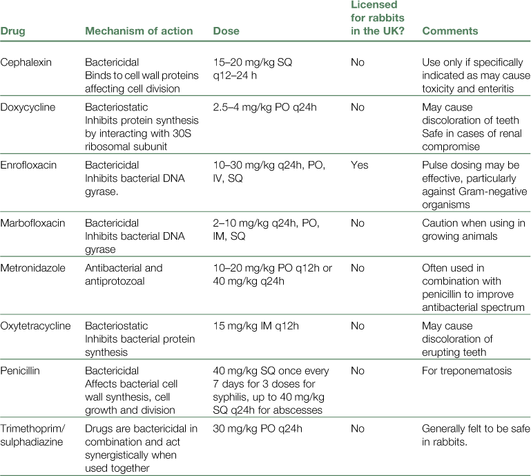
Systemic antibiotics are indicated in severe cases of bacterial skin infections (Table 7.1). Culture and sensitivity may be required. Enrofloxacin or trimethoprim combinations are safe in rabbits and can be used to treat bacterial skin infections. Usually, systemic corticosteroids are not necessary and may be contraindicated due to their immunosuppressive effects (see Section 3.4). If the skin is particularly painful or inflamed, non-steroidal anti-inflammatory drugs (NSAIDs) such as carprofen or meloxicam can be effective. NSAIDs are also beneficial in treating any underlying or painful condition that is restricting grooming.
7.7.1 Moist dermatitis of the dewlap
The skin under the chin and throat can become infected and sore, especially if it is continually wet. Conditions that cause ptyalism increase the risk of moist dermatitis in this region. Dental disease, especially spurs on the cheek teeth, can cause excessive salivation. Saliva runs down the chin and throat, especially on the side of the affected teeth.
Dirty water bottles and damp conditions cause outbreaks of moist dermatitis of the dewlap in rabbits kept under intensive conditions. Some rabbits cannot drink from a water bowl without immersing their dewlap in it. Large breed females are the most susceptible. Substituting sipper bottles for water bowls is recommended as a preventative measure. Licking and chewing the dewlap can be a displacement activity for irritation at another site. For example, obese does that cannot reach their perineum or dorsum may chew their dewlap, which then becomes infected and sore. Removing the primary source of irritation, e.g., perineal dermatitis or cheyletiellosis, is required to cure the dewlap.
Loose-skinned rabbits, especially females, develop huge skin folds under the chin and around the dewlap. These skin folds can easily become infected. Daily cleansing with an antiseptic solution and then thorough drying of the area can be used to control the condition. Alternatively, surgical removal of the skin folds may be required (see Section 7.7.3.1).
7.7.2 Facial dermatitis
Superficial bacterial skin infections around the face can be the result of excessive drooling, fight wounds or chronic epiphora. Epiphora may be due to infection, trauma or blockage of the nasolacrimal duct (see Section 9.6.3). Epiphora is often the first sign of acquired dental disease (see Section 5.6.5). The nasolacrimal duct curls around the apex of the upper primary incisor and becomes blocked by the root if it is elongated (see Figures 9.1 and 9.2). Tears flow down the face instead of down the nasolacrimal duct. The fur under the eye becomes continually wet and mineral deposits combined with sticky mucus cause matting of the fur, encouraging bacterial growth on the skin beneath. Although topical preparations can be helpful in controlling the infection, this condition is difficult to cure (see Section 9.6.3). It is important to keep the area clean and dry. Sedation and clipping of the area may be necessary, allowing the underlying skin to be cleaned and dried. Application of barrier ointment such as petroleum jelly around the eye may be helpful to protect the skin while treatment of the initiating condition is ongoing. Often, the best solution is a bonded companion who will constantly lick and clean this area of the face.
7.7.3 Perineal dermatitis
Perineal dermatitis is usually caused by secondary bacterial infection of the skin around the anus and genitalia. This condition should be viewed as serious and the end result may be myiasis or ‘fly strike’. Grooming difficulties and a fine coat texture often result in mats of fur accumulating in the genital region and on the ventral aspect of the tail, especially when the animal is moulting. These mats of fur absorb urine, adhere to faeces and keep both of these in close apposition to the underlying skin, exacerbating bacterial infection. Areas of inflamed skin can extend down the inside of the thighs to the hocks and are made worse by either urine scalding or contamination of the skin by caecotrophs or both. Uneaten caecotrophs often become entangled in the fur under the tail, especially if they are soft and sticky. Volatile fatty acids in caecotrophs irritate the skin and cause inflammation. There are several conditions that prevent caecotroph ingestion, such as dental disease, obesity, spondylosis, arthritis, sore hocks and inadequate fibre intake (see Figure 8.6). Conditions that can cause urinary incontinence and urine scalding include cystitis, urolithiasis, neurological disease and penile disorders (see Section 12.4.3).
Large perineal skin folds can develop in obese animals and will persist even after the animal has lost weight. Females are more often affected than males. These folds of skin entrap hair, discharges, urine, faeces and necrotic debris. They are prone to chronic infection. Occasionally Psoroptes cuniculi are found in the perineal skin fold of rabbits suffering from ear mite infestation. A crusty exudate forms and mites can be seen on smears taken from the area.
The perineal area is extremely sensitive in rabbits and pain associated with inflamed, infected skin can, in itself, prevent a rabbit from grooming the perineum or ingesting caecotrophs. Lack of grooming starts a vicious circle (see Figure 7.3). Rabbits with painful, infected perineal skin are prone to urethritis. They are reluctant to adopt the correct stance for urination and consequently retain urine and may develop cystitis. Urethritis causes rabbits to dribble urine and dirty wet bedding compounds the situation by scalding the perineal skin. Polydypsic/polyuric rabbits produce copious quantities of urine, which also wets the bedding and exacerbates perineal dermatitis.
Any condition that alters the direction of the jet of urine onto the skin during urination can result in constant urine soaking of the fur and subsequent infection. Conditions of the prepuce or penis, such as scars from fight wounds, can cause penile deviations or partially occlude the urethral opening and alter the direction of urine flow. Rabbits with spinal problems or sore hocks are unable to lift their hindquarters into the correct position to lift their tail and spray urine, which dribbles down the inside of the legs instead (see Figure 12.1).
Treatment of perineal dermatitis is aimed at breaking the vicious circle of inflammation, pain, lack of grooming and secondary bacterial infection. Clipping and cleaning the perineum, treating secondary infection and providing analgesia are effective in most cases. Most rabbits require sedation for the clipping and cleaning, which also offers the opportunity for radiography that may provide a diagnosis. Sludgy urine, urolithiasis, spinal problems or arthritic joints are conditions that can be seen on radiography and predispose to urine scalding or faecal contamination of the perineum. In order to cure perineal dermatitis and prevent recurrence, the underlying reason for faecal or urinary incontinence must be addressed. For example, a high fibre diet may be required to encourage ingestion of caecotrophs or for weight reduction (see Section 1.3.20). NSAIDs are indicated for chronic arthritic conditions such as spondylitis. NSAIDs interfere with prostaglandin synthesis and have the added advantage of reducing caecotroph production (Pairet et al., 1986). Dental diseases that prevent grooming require treatment. Space to exercise and urinate away from the bedding is also necessary.
Rabbits that have developed large folds of skin around the genitalia will benefit from surgical removal of these folds which encourage infection (see Section 7.7.3.1). Entire female rabbits with underlying reproductive disorders that cause vaginal discharges will also need to be spayed. Paraplegic patients nearly always develop a degree of perineal dermatitis and require constant nursing to keep the perineal skin clean, dry and free from matted hair.
7.7.3.1 Perineal dermoplasty
Surgical removal of perineal skin folds is a simple, effective remedy for chronic perineal dermatitis, provided other predisposing factors have been resolved. In some cases it is not possible to cure the underlying cause, but removal of infected skin folds will improve the situation: for example, rabbits with spondylitic problems that are unable to groom properly.
Perineal dermoplasty is performed by making a crescent-shaped incision cranial to the genital opening. The crescent includes the infected and inflamed skin. In male animals, the scrotum and testicles are included so the rabbit is castrated. The amount of skin that needs to be removed varies in each individual. Insufficient skin removal will result in relapse of the condition. Excessive removal will alter the position of the genital orifice and affect the direction of urine flow. The best chance of healing success will be when the edges of the surgical wound are located in an area of skin that is neither infected nor inflamed; however, this is not always possible. There should be no tension on the skin sutures. The incision can be repaired with a soft suture material such as 4/0 Polyglactin 910 (Vicryl Rapide, Ethicon) that does not require suture removal and is comfortable and well tolerated postoperatively. The inflammatory response to polyglactic acid in rabbits is mild (see Section 13.3). Where the surgical incision remains wet postoperatively or is located within an area of infected and inflamed skin, postoperative infection and abscessation is a very real risk. This factor should be considered both in the preoperative planning and the postoperative care plan. Weight-loss advice and support should be offered throughout this time period.
7.8 Fly strike (myiasis)
During the summer months, pet rabbits housed in hutches can be affected by maggot infestation. Healthy rabbits are not affected by fly strike. Obese rabbits are especially prone to the condition.
There is always a reason why the perineal fur becomes contaminated by urine or faeces. Bluebottles (Calliphora) and, more commonly in the UK, greenbottles (Lucilia) are attracted to soiled fur or infected skin to lay their eggs. Primary fly strike usually affects intact skin. The commonest site for fly strike in rabbits is the area at the base of the spine, between the tail and the dorsum. This is a difficult area for rabbits to groom effectively, especially if they are overweight or have a flexibility problem due to spondylosis or arthritis (see Figure 1.19). Dental disease also prevents effective grooming and allows mats to form. Uneaten caecotrophs (see Section 8.6) and damp and dirty bedding both increase the risk of fly strike. Warm temperatures and humidity of 60% or greater promotes the likelihood that fly strike will occur. When the eggs hatch out, the maggots are concealed by matted, soiled fur and may not be obvious until the rabbit becomes unwell. Eggs are laid and hatch within 24 hours, to L1 larvae. These small maggots are non-pathogenic. The moult from L1 to L2 and L3 stages takes 3 days. Both of these stages cause tissue damage. Clinical signs become evident at around 4 days post egg-laying. The skin lesions exude a characteristic smell. Maggots can be intensely irritating for affected rabbits who may become restless and inappetent.
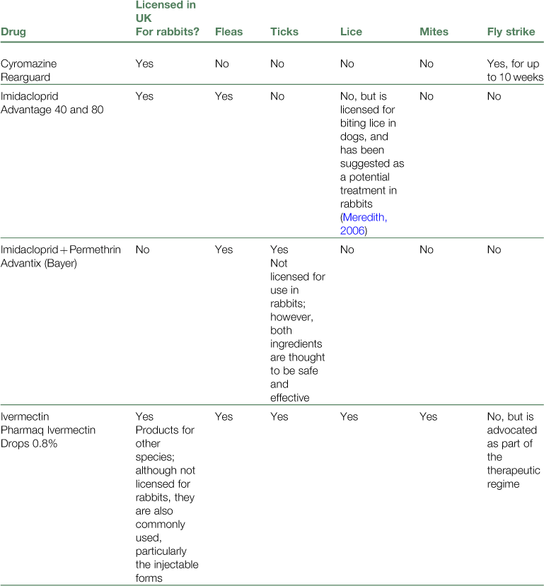
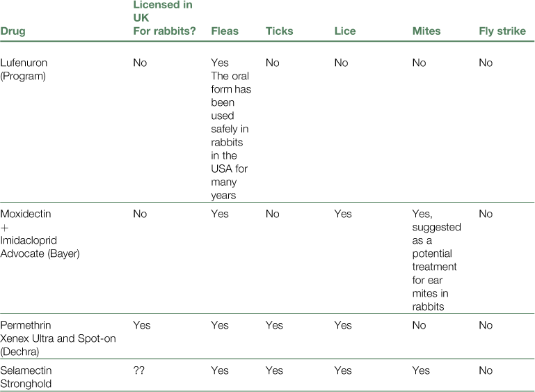
Sedation is usually required to clip away soiled fur from the lesion and pick out the maggots. Fentanyl/fluanisone provides sedation and effective analgesia. All the maggots must be removed from the affected area, which can then be bathed with a medicated shampoo or an antiseptic preparation such as povidone-iodine (Pevidine Surgical Scrub). An insecticidal shampoo can be used, although this is not necessary if all the maggots have been removed. Drying the area with a hair dryer brings out any remaining maggots as they are attracted by the heat; however, care must be taken not to overheat the rabbit or cause thermal damage to the exposed tissue. A precautionary dose of ivermectin should be administered. Antibiotic therapy is indicated; see Table 7.2 for suitable antibiotic choices. An NSAID such as carprofen or meloxicam should also be administered. Inappetent, obese rabbits are at risk of rapidly developing fatal hepatic lipidosis and while all rabbits suffering from fly strike deserve aggressive treatment, obese patients are more vulnerable to the potentially fatal side effects of anorexia. Fluid therapy, syringe feeding, analgesia and motility stimulants are necessary. Addressing the underlying cause of perineal soiling is required to prevent further episodes of fly strike, although providing some type of fly screen and ensuring a clean dry bed are beneficial. Prophylactic insecticidal preparations such as Rearguard (Novartis Animal Health) and Stronghold (Pfizer) are licensed for this use in rabbits and may be used but such preparations do not address the underlying cause. Fipronil (Frontline) is not advisable, as rabbits can suffer adverse reactions that may be potentially fatal.
In the USA, rabbits can be affected by a warble fly, Cuterebra cuniculi. The incidence of infestation decreases with age, which correlates with the development of immediate and delayed-type hypersensitivity reactions to the larvae. Initial lesions include subcutaneous cystic structures. As the larvae enlarge, a breathing hole or fistula develops. The surrounding coat becomes moist and matted and secondary bacterial infection is common. The lesions are often painful, and significant abscessation may be associated with the lesion. Treatment consists of surgical removal of the larvae without crushing or damaging them, as well as any associated abscessation (see Chapter 6). The condition is prevented by eliminating contact with the warble fly (Scott et al., 1995).


