Chapter 3 Skeletal system
The skeletal system is the ‘framework’ upon which the body is built – it provides support, protection and enables the animal to move (Fig. 3.1). The joints are considered to be an integral part of the skeleton. The skeletal system is made of the specialised connective tissues, bone and cartilage.
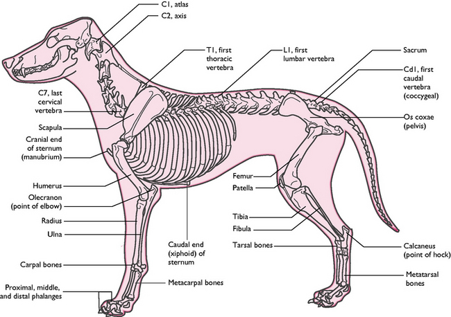
Fig. 3.1 The skeleton of the dog showing the major bones.
(With permission from Colville T, Bassett JM 2001 Clinical anatomy and physiology for veterinary technicians. Mosby, St Louis, MO, p 102.)
The functions of the skeletal system are:
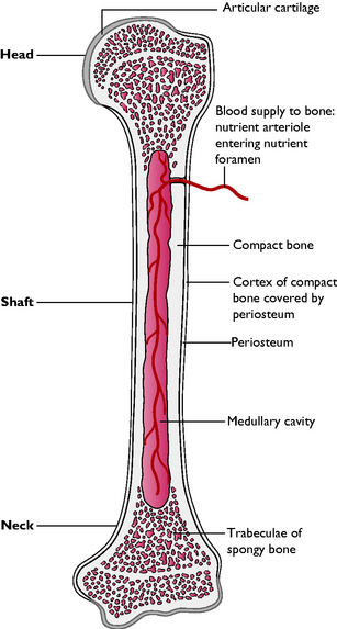
Bone structure and function
Bone shape
Bones can be categorised according to their shape:
Some specialised types of bone are:
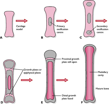
Development of bone
Endochondral ossification
The process of endochondral ossification (Fig. 3.3) is as follows:
The skeleton
The skeleton (Fig. 3.1) can be divided into three parts.
When studying the anatomy of the skeletal system it is helpful to understand the terms that are used to describe the various projections, passages and depressions that are found on and within bones.
The axial skeleton
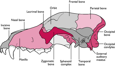
The skull
Cranium
The caudal part of the skull that provides the bony ‘case’ in which the brain sits is called the cranium (Figs 3.4, 3.5). The bones of the cranium include:
Nasal chambers
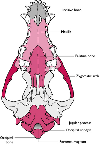
The most rostral part of the skull carries the nasal chamber, the sides of which are formed by the maxilla and the roof by the nasal bone. The nasal chamber is divided lengthways into two by a cartilaginous plate called the nasal septum. Each of the chambers is filled with delicate scrolls of bone called the nasal turbinates or conchae. These are covered in ciliated mucous epithelium (see Ch. 8). At the back of the nasal chamber, forming a boundary between the nasal and cranial cavities, is the ethmoid bone. In the centre of this bone is the cribriform plate – a sieve-like area perforated by numerous foramina through which the olfactory nerves pass from the nasal mucosa to the olfactory bulbs of the brain (see Ch. 5).
The roof of the mouth is called the hard palate and is formed from three bones on the ventral aspect of the skull: the incisive bone or premaxilla, part of the maxilla and the palatine. The incisive bone is the most rostral and carries the incisor teeth (Fig. 3.5).
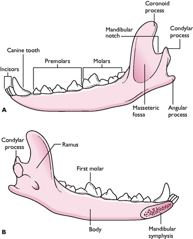
Mandible
The mandible or lower jaw is comprised of two halves or dentaries, joined together at the chin by a cartilaginous joint called the mandibular symphysis. Each half is divided into a horizontal part, the body, and a vertical part, the ramus (Fig. 3.6). The body carries the sockets or alveoli for the teeth of the lower jaw. The ramus articulates with the rest of the skull at the temporomandibular joint via a projection called the condylar process. A rounded coronoid process, which projects from the ramus into the temporal fossa, is the point to which the temporalis muscle attaches (Fig. 3.6). There is a depression on the lateral surface of the ramus, the masseteric fossa, in which the masseter muscle lies.
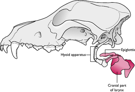
Hyoid apparatus
The hyoid apparatus lies in the intermandibular space and consists of a number of fine bones and cartilages joined together in an arrangement that resembles a trapeze (Fig. 3.7). The hyoid apparatus is the means by which the larynx and tongue are suspended from the skull. The apparatus articulates with the temporal region of the skull in a cartilaginous joint.
< div class='tao-gold-member'>
Stay updated, free articles. Join our Telegram channel

Full access? Get Clinical Tree


