Skeletal Muscle and Lameness

Chapter 83Skeletal Muscle and Lameness


Diagnosis of Specific Muscle Disorders in the Horse


Diagnosis of a particular muscle disorder is best accomplished with a thorough neuromuscular examination. The key components of the examination include the following.


Ancillary Diagnostic Tests


Muscle Enzymes


Skeletal muscle necrosis may be identified by determining the activity in blood of serum enzymes or proteins that are normally present in high concentration within intact muscle cells but leak out into the bloodstream following cell damage. Three enzymes are used routinely to assess muscle necrosis: creatine kinase (CK), aspartate transaminase (AST), and lactate dehydrogenase (LDH). Serum myoglobin has also been used as a marker of acute muscle necrosis.2,3 The permeability of the muscle cell membrane, rate of enzyme production, alternate tissue sources of the enzyme, and rate of enzyme excretion/degradation may also influence serum enzyme activities.


Nuclear Scintigraphy


Nuclear scintigraphy is useful for identification of some forms of muscle damage and may alert the clinician to an area of deep muscle damage that had not been suspected based on clinical examination. In human athletes, technetium-99m stannous pyrophosphate has been used to assess the degree of skeletal muscle damage and to delineate areas of damage.14,15 It is thought that abnormal uptake of the radiopharmaceutical reflects an early stage of muscle damage from episodic ischemia, which is reversible in some fibers but may lead to muscle necrosis in others.14


Technetium-99m–methylene diphosphonate (MDP) is taken up in some damaged muscle in the horse and is best seen in the bone (delayed) phase images, that is, 3 hours after injection. Scintigraphy has been used most commonly in horses with a history of poor performance, with or without stiffness after exercise, to confirm a diagnosis of equine rhabdomyolysis.16 The mechanism of MDP binding is unknown, but the release of large amounts of calcium from damaged muscle or the exposure of calcium binding sites on protein macromolecules in the damaged muscle may be responsible. Diffuse linear areas of increased radiopharmaceutical uptake (IRU) are commonly seen in the caudal epaxial muscles and the muscles of the hindquarters and thigh in some but not all horses with ER (Figure 83-5). Less commonly there is IRU in the triceps and latissimus dorsi muscles.


The use of scintigraphy for the diagnosis of other muscle injuries has not been documented in the horse, but in one author’s experience (SJD) it can be helpful in some horses with either proximal forelimb or hindlimb muscle injuries. Uptake of the radiopharmaceutical tends to be much more focal and much less intense than in horses with rhabdomyolysis. In some, but not all, horses the region of IRU has correlated with a region of increased echogenicity identified ultrasonographically.


Muscle Biopsy


The routine examination of muscle biopsies has resulted in the identification of a number of specific equine myopathies. To fully characterize a neuromuscular disorder and its rate of progression, muscle fiber sizes, shapes, and fiber type distribution, mitochondrial distribution, polysaccharide staining pattern, neuromuscular junctions, nerve branches, connective tissue, and blood vessels should be examined in frozen sections using a battery of tinctorial and histochemical stains.19


A number of basic pathological responses of muscle can be identified in formalin-fixed, paraffin-embedded sections. These include inflammation, muscle fiber necrosis, muscle fiber regeneration, variations in muscle fiber sizes and shapes, alterations in the number of cell nuclei, vacuolar change, and proliferation of connective tissue. However, there are many pathological alterations that cannot be detected in formalin-fixed tissue but can readily be seen in histochemical stains of fresh-frozen biopsy samples.20 Histochemical stains of frozen tissue allow muscle fiber types to be distinguished, differentiation between neurogenic and myogenic atrophy, characterization of vacuolar storage material, characterization of inclusion bodies, and assessment of mitochondrial density. In addition, frozen samples may be used for biochemical analysis of substrate concentrations and enzyme activities, as well as DNA isolation.


When considering collection of muscle biopsies, some general guidelines are applicable. Preferably, samples should be collected from what is considered abnormal/diseased muscle. A 6-mm outer diameter (Jorgen KRUUSE A/S, Langeskov, Denmark) percutaneous needle biopsy technique can be used to obtain small muscle samples through a 1.5-cm skin incision using a local anesthetic solution subcutaneously. If this technique is used, enough muscle should be obtained to form a 1.5-cm2 sample at a minimum. However, these samples do not tolerate well shipment to an outside laboratory. The optimum biopsy for shipment of histopathological tissues to a laboratory is collected using surgical or open techniques and performed under local analgesia. Care must be exercised to infiltrate only the subcutaneous tissues, not the muscle, with the local anesthetic solution. The objective is to obtain approximately 2-cm3 of tissue; hence a suitably long skin incision is required. Subsequently two parallel incisions 2 cm apart should be made longitudinal to the muscle fibers with a scalpel. The muscle should only be handled in one corner using forceps, and care should be taken not to crush the tissue. The muscle sample is then excised by transverse incisions 2 cm apart, and the tissue is fixed appropriately.


Samples submitted for routine histopathology can be placed in formalin. Samples for histochemical analysis require fixation in isopentane (methylbutane) chilled in liquid nitrogen to ensure rapid freezing and minimization of freeze artifact. In the field, where freezing is not possible, fresh samples wrapped in gauze slightly moistened with saline can be shipped in a water-tight hard container on icepacks to specialized laboratories. Samples that potentially may be used for biochemical analysis should be immediately frozen in liquid nitrogen. Samples for electron microscopy (EM) require appropriate fixation in glutaraldehyde preparations. Ideally, thin sections of muscle for EM should be clamped in vivo to maintain fibers at a resting length before they are excised. However, if pathology other than the alignment of thick and thin myofilaments is to be investigated, small muscle pieces can be excised and placed directly in appropriate EM fixative.


Responses of strips of fresh muscle to stimuli such as caffeine, halothane, and a variety of other agents can be performed on site by specialized laboratories, but these tests are largely research tools.21,22


Electromyography


A specific diagnosis of the cause of muscle atrophy, muscle fasciculations, or myotonic dimpling after tapping the muscle can be aided by performing electromyography (EMG). EMG of normal skeletal muscle shows a brief burst of electrical activity when the needle is inserted in muscle and then quiescence, unless motor units are recruited (motor unit action potentials), or the needle is very close to a motor endplate (miniature endplate potentials). Normal muscle shows little spontaneous electrical activity unless the muscle contracts or the horse moves. Motor unit action potentials can be evaluated to assess amplitude, duration, phase, and number of phases. Myopathic changes include a decrease in duration and amplitude of motor unit action potentials.23,24 Horses with abnormalities in the electrical conduction system of muscle, or denervation of motor units, show abnormal spontaneous electrical activity in the form of fibrillation potentials, positive sharp waves, myotonic discharges, or complex repetitive discharges.


Based on the information obtained on neuromuscular examination and muscle biopsy, a diagnosis can usually be obtained. The following classification system may be helpful to narrow down rule-outs for muscle disease in horses:


Muscular Pain, Strain, and Tears


The role of muscle pain and injury in lameness and poor performance in the horse is rather poorly recognized. In human athletes, muscle fatigue, muscle stiffness, and muscle soreness are well-recognized entities, although the pathological processes in the absence of detectable structural abnormalities are not completely understood. Increased intramuscular pressure may be associated with muscle pain after prolonged vigorous exercise in human athletes.14


Delayed-onset muscular stiffness or soreness (DOMS) is recognized in people as pain that develops 24 to 48 hours after unaccustomed use of certain muscles and usually resolves spontaneously, assuming the muscles are not overworked again.25 Continued overstress may result in structural damage to myofilaments. However, specific training involving the activity that provoked the original DOMS decreases the amount of soreness associated with that condition over time.


Muscle soreness in the pectoral region after repeated jumping efforts is commonly recognized, especially in event horses several hours after completing the cross-country phase of a Three Day Event.26 It seems to improve with massage.


Muscle fiber tearing and hemorrhage can result in acute muscular pain in human athletes. A palpable defect or swelling can be detected in superficial muscles. For deeper muscles, ultrasonography is required for accurate diagnosis.


Muscle fibrosis and mineralization have been well documented in the horse following tearing of the semimembranosus and semitendinosus muscles (see Fibrotic Myopathy section, page 558), but acute lesions here and elsewhere in the limbs have been poorly documented.27-29 The use of diagnostic ultrasonography17,18 has helped in the diagnosis of both acute and more chronic muscle lesions, but diagnosis often remains a challenge because of the deep location of some affected muscles and the lack of localizing clinical signs.


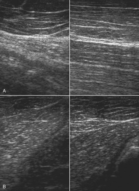
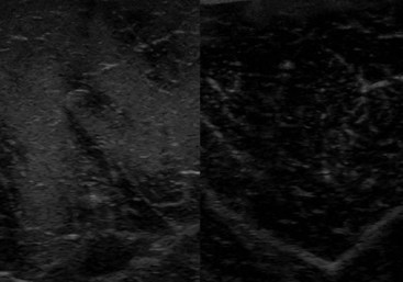
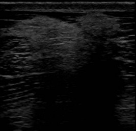
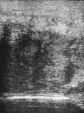
In one author’s (SJD) experience, the most commonly recognized muscle injury sites in the forelimb include biceps brachii, brachiocephalicus, the pectorals, and the musculotendonous junction of the superficial digital flexor (Figures 83-1 to 83-4). In the hindlimb, semimembranosus and semitendinosus, adductor, gracilis, gluteal, and gastrocnemius muscle injuries have been recognized most frequently. Acute muscle tearing and hemorrhage can result in severe pain and lameness and other clinical signs mimicking colic. Swelling around the damaged muscle, assuming it is superficial, may not appear until 24 to 48 hours later.


Muscle tension and spasm in the thoracolumbar region are well-documented sources of pain contributing to poor performance in association with primary hindlimb lameness, but primary muscle pain in this region has often tended to be overlooked by many veterinarians, although recognized by physiotherapists. Localized muscle soreness and the interpretation of abnormal sensitivity of acupuncture points are potentially confusing. Protective muscle spasm may also develop secondary to a primary lesion of either the thoracolumbar spine or the sacroiliac region (see Chapters 49 to 52).


Jeffcott et al30 demonstrated that injection of lactic acid into the left longissimus dorsi muscles could significantly diminish performance of Standardbred (STB) trotters worked at speed on a treadmill, although changes in gait were subtle.


One author (SJD) has seen a number of horses that had suddenly lost performance during competition or training, either following a particularly extravagant jump or after an awkward jump. The horses had subsequently become reluctant either to jump or to gallop downhill. Clinical examination revealed intense muscle spasm in the caudal thoracic and lumbar regions, with associated pain. Manipulation to release muscle spasm resulted in relief of pain and rapid restoration of normal performance. Acute back muscle pain may also be induced by a fall.


Localized back muscle soreness is readily induced by a poorly fitting saddle. It may also be caused by a rider who either sits crookedly or is unable to ride completely in balance with the horse. This may be because of the ineptitude of the rider or the shape of the saddle and the way in which it sits on a particular horse and thus positions the rider. Such muscle pain is usually localized to the saddle area and may be associated with slight soft tissue swelling. Thermographic examination may be helpful to demonstrate to an owner the associated localized inflammation. Pressure measurements can also be used (see Figure 117-6), although there is some variability in the accuracy of different commercially available systems.


Diagnosis


History and Clinical Examination


It is important to establish whether there was a history of a fall or other traumatic event, the duration of clinical signs, whether swelling was noted, and whether the horse had exhibited lameness or had performed poorly.


The detection of muscle swelling from recent trauma or loss of muscle bulk as a result of fibrosis, chronic injury, or atrophy requires the horse to stand completely squarely, bearing weight evenly on all limbs and looking straight ahead. The horse should be appraised visually and by careful, systematic palpation, looking for defects in the muscle, muscle swelling, areas of abnormal muscle firmness from fibrosis, muscle tension, or spasm, and areas of pain.


In an acute injury resulting in muscle tearing or rupture, it may be possible to palpate a defect in the very early stages, but this will become filled with hemorrhage, inflammatory exudate, and edema. Careful palpation should enable detection of most acute superficial muscle injuries, but localization of deeper muscle injury may be more difficult. Identification of chronic muscle strain is more challenging because clinical signs are more subtle. The horse must be as relaxed as possible to assess properly the response to firm and deep palpation. If the muscle is sore, the horse may react by increasing tension in anticipation of pain or by pulling away. There may be “knots” within the area of damaged muscle.


The neck, limbs, and thoracolumbar region should be moved passively to detect any limitations in movement or pain induced by movement. The horse should be observed moving at both the walk and the trot to identify any characteristic gait abnormalities, such as an abnormal hind foot placement from fibrotic myopathy (see Chapter 48) or sinking of a front fetlock as a result of rupture at the musculotendonous junction of the superficial digital flexor (see Chapter 13)image. However, it must be borne in mind that muscle soreness resulting in compromise of performance may not result in overt lameness because pain may only be induced when the muscle contracts strongly or is stretched maximally. Pain associated with a brachiocephalicus muscle in a dressage horse may only be evident in particular movements such as half pass (see Figure 83-3). A show jumper with sore gluteal muscles may not push off as strongly with the affected limb, resulting in the hindquarters drifting toward the ipsilateral side as the horse jumps.


Exertional Rhabdomyolysis


ER has numerous etiologies and is a common complex cause of poor performance. About 3% of exercising horses had an episode of ER in the past 12 months.37 It occurs in a variety of breeds, including draft breeds, Warmbloods, Thoroughbreds (TB), STBs, Arabians, Morgans, Quarter Horses, Appaloosa, American Paint horses, and many more.37-41 In draft breeds, ER can be particularly debilitating, and terms such as Monday morning disease, azoturia, or paralytic myoglobinuria are used.42 A milder syndrome occurs in lighter breeds, and the terms tying-up, set fast, myositis, and chronic intermittent rhabdomyolysis are used to describe muscle necrosis or muscle cell stress following any form of exercise in the lighter horse breeds.43,44 Several specific causes have been identified for ER. Otherwise successful athletic horses may have a sporadic episode of ER from extrinsic causes such as dietary imbalances, concurrent respiratory infections, and inappropriate training regimens. In horses chronically afflicted with ER there may be an intrinsic dysfunction of muscle metabolism or muscle contraction.


Sporadic Exertional Rhabdomyolysis


The most common extrinsic cause of sporadic ER is exercise that exceeds the horse’s underlying state of training.44 Horses that are advanced too quickly in training, horses that are only ridden sporadically while being continually fed full rations, and horses performing strenuous exercise such as racing or endurance riding without sufficient conditioning commonly develop rhabdomyolysis. In addition, rhabdomyolysis may be more common in horses exercising during an outbreak of respiratory disease. Both equine herpesvirus–1 and equine influenza virus have been implicated as causative agents.38,45


Treatment of Acute Rhabdomyolysis


If an attack has occurred during exercise some distance from where the horse is normally stabled, the horse should not be made to walk home. It should be transported back home or left in a nearby stable. If an attack has occurred at a competition, the horse should be treated there and should not be transported home over a long distance until at least 24 to 48 hours later.


The objectives of treatment are to relieve anxiety and muscle pain, as well as to correct fluid and acid-base deficits. Acetylpromazine (0.04 to 0.07 mg/kg), an α-adrenergic antagonist, is helpful in relieving anxiety and may increase muscle blood flow. Its use is contraindicated in dehydrated horses. Alternatively, xylazine (0.4 to 0.8 mg/kg) may provide short-term relief from anxiety. In horses with extreme pain, detomidine (0.02 to 0.04 mcg/kg) combined with butorphanol (0.01 to 0.04 mg/kg) provides excellent sedation and analgesia. Nonsteroidal antiinflammatory drugs (NSAIDs) such as ketoprofen (2.2 mg/kg), phenylbutazone (2.2 to 4.4 mg/kg), or flunixin meglumine (1.1 mg/kg) provide additional pain relief. Analgesic treatment is continued to effect, but most horses are relatively pain free within 18 to 24 hours.


Intravenous or intragastric dimethyl sulfoxide (as a <20% solution) can be used as an antioxidant, antiinflammatory, and osmotic diuretic in severely affected horses. Corticosteroid administration is advocated by some veterinarians in the acute stage. If the horse is recumbent, methyl prednisolone succinate (2 to 4 mg/kg intravenously [IV]) should be given once. Muscle relaxants such as methocarbamol (5 to 22 mg/kg, IV slowly) seem to produce variable results, possibly depending on the dosage used. The administration of dantrium sodium (2 to 4 mg/kg orally [PO]) in severely affected horses may decrease muscle contractures and possibly prevent further activation of muscle necrosis. This can be repeated in 4 to 6 hours.


Severe ER can lead to renal compromise from ischemia and the combined nephrotoxic effects of myoglobinuria, dehydration, and NSAIDs. The first priority in horses with hemoconcentration, or myoglobinuria, is to reestablish fluid balance and induce diuresis. In horses with mild rhabdomyolysis, administration of fluids via a nasogastric tube may be adequate, but generally fluids are better given intravenously. Balanced polyionic electrolyte solutions are best. If severe ER is present, then isotonic saline or 2.5% dextrose in 0.45% saline may be necessary because horses often have hyponatremia, hypochloremia, and hyperkalemia. If hypocalcemia is present, then supplementing intravenous fluids with 100 to 200 mL of 24% calcium borogluconate is recommended, but serum calcium should not exceed a low normal range. Affected horses are usually alkalotic, making bicarbonate therapy inappropriate.48


Ten liters of fluids may be given rapidly. The total fluid replacement is based on an estimation of the degree of dehydration and the clinical response: if the horse is mildly dehydrated (5%), give 10 L fast and then 15 L over the next 4 to 6 hours; if dehydration is severe (20%), give 10 L fast and then 50 L at 4 L/h. If the horse is recumbent, consider using at least two intravenous giving sets and infusing into both the jugular and the cephalic veins. Suture the catheters in place.


Ideally, reassessment of the packed cell volume and concentrations of total plasma protein and serum electrolytes after the initial period of therapy should provide a successful guide for the therapeutic regimen. However, in the practical situation, the clinical response to therapy is usually an adequate indicator. In severely affected horses, regular monitoring of blood urea nitrogen and/or serum creatinine is advised to assess the extent of renal damage. Diuretics are usually contraindicated unless the horse is in oliguric renal failure.


Horses should be stall rested on a hay diet for a few days. Small paddock turnout in a quiet area for a few hours twice a day is then helpful. Horses may be handwalked at this time but not for more than 5 to 10 minutes at a time. For horses with extrinsic (sporadic) ER, rest with regular access to a paddock should continue until serum muscle enzyme activities are normal. Training should be resumed gradually, and a regular exercise schedule that matches the degree of exertion to the horse’s underlying state of training should be established. Avoid lunging exercise until the horse is back in normal work. If the horse has a day or several days off, the dietary energy concentrations should be reduced accordingly.


Chronic Exertional Rhabdomyolysis


Horses that have repeated episodes of ER from a young age, or from the time of purchase, or when they are put back into training after a long period of rest may have a chronic dietary imbalance or underlying intrinsic abnormality of muscle function. Chronic forms of ER are seen in many breeds of horses, including draft horses, Warmbloods, Quarter Horses, American Paint horses, Appaloosas, TBs, Arabians, STBs, Morgans, and crossbreds.39,44,49-55 Many of the horses with intrinsic muscle defects will have repeated episodes of rhabdomyolysis with minimal exercise, even when the dietary and training recommendations for sporadic ER are followed. Three specific intrinsic causes of ER have been identified to date: recurrent exertional rhabdomyolysis (RER),56 type 1 polysaccharide storage myopathy (PSSM),57,58 and type 2 PSSM.59 It is likely that there are other specific causes that have yet to be identified (idiopathic chronic ER). In all these intrinsic forms of chronic ER, it appears that there are specific environmental stimuli that are necessary to trigger muscle necrosis in genetically susceptible animals.46,56 Horses cannot be cured of a susceptibility to this condition, but if the specific disease is identified, changes in management can be implemented to minimize episodes of rhabdomyolysis.


Jun 4, 2016 | Posted by in EQUINE MEDICINE | Comments Off on Skeletal Muscle and Lameness

Full access? Get Clinical Tree

Get Clinical Tree app for offline access