Seizure Disorders: Narcolepsy

CHAPTER 18 Seizure Disorders


Narcolepsy


DEFINITION


See Video 18-1 to introduce yourself to the subject of seizure disorders and so you can better appreciate the following discussion. This video shows Victory Forth, a 9-year-old female quarter horse, that has had a series of generalized seizures for the first few weeks after her last three foalings. Two seizures are shown on the video between which she was normal for approximately 1 hour.


Remember that a neuron is an excitable tissue constantly held in check. When this constant inhibition is interrupted, a seizure may be the result! Neurons communicate with each other via excitatory and inhibitory connections. Normal neuronal activity represents a balance of these excitatory and inhibitory influences. When imbalance results in excessive excitation, abnormal neuronal activity occurs and is manifested as a seizure. A seizure represents the uncontrolled synchronous discharge of neurons that is initiated in the prosencephalon. It is usually a brief episode as the brain usually rapidly corrects the imbalance in these neuronal influences. A seizure is the most common clinical complaint that involves the nervous system that a practicing veterinarian will encounter.


The terms seizure, convulsion, epilepsy, and fit are synonyms for a brain disorder expressed as a paroxysmal transitory disturbance of brain function that has a sudden onset, ceases spontaneously, has a tendency to recur, and originates in the prosencephalon. The term epilepsy is usually used for seizures that are recurrent. Those of unknown cause are called idiopathic epilepsy. The initial seizure focus may involve only a small number of highly unstable neurons that spontaneously discharge. This first episode may induce surrounding neurons to discharge, resulting in a progressive spread, or generalization, of the seizure. The pathogenesis of seizures has been and still is currently the subject of extensive investigation.*


TERMINOLOGY


The period of the seizure is known as the ictus, or the attack. The manifestation of the seizure is variable. Any unusual involuntary phenomenon that is episodic and recurrent in nature should be evaluated as a seizure disorder. Such phenomena include loss or derangement of consciousness, excessive or decreased voluntary muscle tone or movement, visceral muscle activity, and altered behavior.


Postictal depression refers to the period of recovery after a seizure when the patient may wander around in a state of confusion, may propulsively walk in circles or bump into objects because of its central blindness, or may sleep for a prolonged period or exhibit hyperphagia. This phase has the appearance of a patient whose neurons are exhausted from the excessive activity of the seizure. The length and form of postictal depression are variable. No correlation exists between the severity and length of the seizure and the severity, duration, or nature of the postictal phase. A short partial seizure may be followed by a longer, more complex postictus than a generalized seizure. As a rule, the postictal phase lasts less than an hour, but much longer periods of up to 1 to 2 days are possible. In horses, the postictal phase may last for 3 to 4 days.


The interictal period is the period between seizures after the patient has recovered from the postictal period. This phase is when the neurologic examination should be performed.


An isolated seizure is a seizure that occurs only once in a 24-hour period. Cluster seizures are when two or more seizures occur in 24 hours separated by normal interictal periods. Status epilepticus is when the seizure state is continual for more than 5 minutes or when a series of seizures occur without full recovery of consciousness between the seizures for 30 minutes.5,93 The seizure is continually repeated with no interictal period. Cluster seizures and status epilepticus are medical emergencies.


Descriptions of seizures usually include the terms aura and prodrome. The prodrome or prodromal period precedes the seizure and consists of a change in the sensorium of the patient that is exhibited in its behavior. The aura is defined as the initial focal motor or sensory signs that may precede a generalized seizure by a few seconds. If the aura is observed, it may suggest the site of the seizure focus. We do not regularly use these terms because they are often difficult to recognize in our patients. However, some owners will report recognizing a change in their pet’s behavior, a prodrome or aura, that is predictive of a seizure.


CLASSIFICATION


The classification of seizures in animals is controversial and cannot be directly correlated with that used in humans.80 We believe the following discussion best serves the experience we have had with seizures in domestic animals, which are most common in the canine species. Seizures can be classified as focal, partial, or general; those that begin as partial seizures may subsequently generalize.


A focal seizure is a nonclinical spontaneous discharge of a small group of prosencephalic neurons without any spread. It can be observed only on an electroencephalogram (EEG) and should not be confused with a partial seizure, which is a clinical entity. Focal seizures are sometimes present in the interictal period of dogs diagnosed as idiopathic epileptics. Notably, some clinicians use the terms focal and partial as synonyms, which is technically incorrect.


A partial seizure is a focal seizure that has a limited spread and is observed clinically. The nature of the clinical signs will reflect the area of the prosencephalon where the seizure focus is located and consists of varying degrees of abnormal motor and sensory behavior with or without loss of consciousness. Lateralizing clinical signs often indicate which side of the prosencephalon is affected. Most partial seizures occur in animals that have a structural lesion in the prosencephalon. Occasionally, partial seizures are idiopathic. Two categories are described for partial seizures.


1 A simple partial seizure reflects primarily abnormal motor neuron discharge with no disturbance of the patient’s sensorium. Episodic tremors, head turning, limb flexion, facial muscle twitches, head and neck sporadic myoclonus, sialosis, and mydriasis are examples. In Chapter 8, see Videos 8-29 through 8-31 for examples of sporadic myoclonus, which is a form of simple partial seizure. Some of these simple partial seizures may also be difficult to differentiate from a movement disorder, which we describe in Chapter 8. A simple partial seizure that involves only the limbs or face on one side of the body is considered as a lateralizing clinical sign and indicates that the seizure focus or lesion is in the opposite cerebral hemisphere. For most simple partial seizures, the seizure focus is thought to be in the motor area of the cerebral hemisphere. In humans, a simple partial (focal motor) seizure that consists of an initial tonic contraction followed by clonic contractions of one hand that spreads to the rest of that forelimb, the ipsilateral face, and pelvic limb is called a Jacksonian seizure. This type of seizure indicates a focus in the contralateral motor cortex.

2 A complex partial seizure includes some disturbance of the patient’s sensorium, which is expressed as a behavioral abnormality.33 Some examples are episodes of staring into space, maniacal running, tail chasing, flank attacking, fly or light biting, abnormal aggression or rage, and just brief episodes of loss of consciousness. These complex partial seizures are often called psychomotor seizures because of the behavioral component. The presence of abnormal behavior in the seizure suggests that the seizure focus includes involvement of the limbic system. A complex partial seizure may terminate in a generalized seizure.

A generalized seizure is the most common form that occurs in domestic animals and affects the brain diffusely. This form of seizure has been called a grand mal seizure. The petit mal seizures that are described in humans consist of brief lapses in the conscious state with a specific EEG pattern. These types of seizures have not been recognized in domestic animals. A generalized seizure is the form of seizure seen in the 9-year-old Quarter horse in the introductory video (Video 18-1) for this chapter. The initial seizure focus may be in one cerebral hemisphere that immediately spreads to the thalamus, which diffusely activates the entire cerebrum through its neurons that function in the diffuse cortical projection system, or the seizure may originate in this thalamic system. The result is a loss of the conscious state by the patient, a tonic contraction of most of the antigravity skeletal muscles, recumbency followed by periods of tonic muscle activity, and clonic muscle activity, which causes running-like movements of the limbs. Jaw clinching or chewing movements may occur along with sialosis, dilated pupils, hair erection, and sometimes urinary and fecal excretions. Apnea may occur during a tonic phase of the seizure. This form of seizure usually lasts from 30 seconds to 3 minutes, followed by a variable period of postictal clinical signs and then complete recovery.


PATHOGENESIS


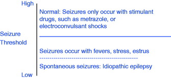
To understand the basis for seizures, consider that neurons have what we call a seizure threshold. This neuronal threshold is determined by their environment, which is genetically determined. Seizures result when this neuronal environment is disturbed and the threshold is lowered. Most commonly, the seizure originates in disturbed prosencephalic neurons. Consider this neuronal threshold in the following simple graph diagram.


image


What comprises this neuronal environment? This concept is obviously very complex, but at the least it includes (1) the structure of the dendritic zones and all their synapses, as well as those on the neuronal cell body; (2) the neuronal lipoprotein cell membrane, including the plethora of ion channels that are influenced by the neurotransmitters and the enzymes involved with their activity, such as sodium-potassium-adenosine triphosphatase; (3) the ionic environment of the neurons that includes the availability of sodium, chloride, calcium, and potassium; and (4) the concentration of neurotransmitters that includes those concerned with excitation—glutamate, aspartate, and acetylcholine—and those concerned with inhibition—gamma aminobutyric acid, glycine, taurine, and norepinephrine. In addition, this neuronal environment includes the adjacent neurons and the astrocytes, which also have synapses with neurons and other astrocytes. Astrocytes regulate the transfer of metabolites and ions through the blood vessel walls and have a role in metabolizing many of the neurotransmitters.73Fig. 18-1 will help you appreciate the anatomic background of this neuronal environment. Alteration of any one or more of these components may lower the neuronal threshold enough to precipitate a seizure. These alterations comprise the differential diagnosis for seizures.
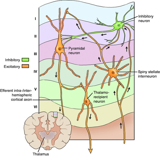


Intracranial Disorders


Intracranial in this discussion refers to structural disorders.101 A thorough neurologic examination will often reveal one or more neurologic deficits in the interictal examination, which indicates a structural lesion.


1 Malformation. Many of the malformations described in Chapter 3 that involved the prosencephalon are possible causes of seizures. Obstructive hydrocephalus is the most common. Lissencephaly is another cause, with the onset of seizures at a few years of age. Hydranencephaly is rare in dogs, but we have seen a miniature poodle with unilateral hydranencephaly that began to have seizures at a few months of age. Abnormalities in neuronal migration that cause various cerebrocortical dysplasias are relatively subtle malformations that are more difficult to identify. Considerable interest exists in these cortical dysplasias being a cause of seizures in some dogs diagnosed with idiopathic epilepsy.


3 Neoplasia. In our experience, neoplasms are the most common structural cause of seizures, as well as the most common lesion to cause seizures initially and no other clinical neurologic deficit.16,100,102 Never exclude a brain neoplasm or other structural lesion because the interictal neurologic examination is normal. Lesions can occur in large areas of cerebrum that we cannot recognize on the clinical neurologic examination. For example, we often see large neoplasms of the olfactory bulbs and frontal lobes in dogs; these neoplasms cause seizures without any apparent neurologic abnormalities on our neurologic examination. Primary prosencephalic neoplasms (neurectodermal) are much more common than metastatic neoplasms, and they are more common in dogs and cats than other species of domestic animals. In cats, meningiomas are the most common brain neoplasm. However, in our experience, brain neoplasms in cats, especially meningiomas, do not seem to cause seizures as frequently as they do in dogs.


5 Degeneration. A wide variety of disorders cause various forms of neuronal degeneration in the prosencephalon.51 Vascular compromise, cerebrovascular accident, and stroke are all terms related to varying degrees of ischemia or infarction of the brain that, when they occur in the prosencephalon, are possible causes of a seizure. These lesions include those associated with Cuterebra spp. myiasis in cats, hypertensive vasculopathy most commonly associated with chronic renal disease, or hyperthyroidism in cats and chronic hypothyroidism in dogs associated with the rare occurrence of cholesterol plaque formation in the walls of cerebral blood vessels. The neonatal encephalopathy of foals is another example.79 Other degenerations include thiamin deficiency encephalopathy in cats and ruminants, osmolar disorders related to excessive salt or water ingestion, and lead intoxication, as well as other toxicities that include organophosphates, chlorinated hydrocarbons, the artificial sweetener xylitol, caffeine in chocolate, and ethylene glycol.48,131 Metabolic disorders such as the inherited storage diseases and mitochondrial encephalopathies often cause seizures, as does heat stroke.21


Extracranial Disorders


Extracranial causes of seizures are disorders that affect primarily other body systems that, in turn, affect the metabolism of neurons in the central nervous system (CNS). The interictal neurologic examination is usually normal in these patients. However, the physical examination may reflect a systemic abnormality that is related to the cause of the seizures. Some overlap exists with the intracranial group because some of these disorders cause degenerative lesions in the prosencephalon. This group emphasizes what needs to be considered when you study the history, do your physical examination, and order ancillary studies.


l Hypoglycemia. Hypoglycemia from any origin is a common cause of seizures.69 The most common cause is a functional neoplasm of the beta cell in the pancreatic islet that produces insulin.14,23,25,106 The common but inappropriate term for this neoplasm is insulinoma, which, by its derivation, means a swelling of insulin. My younger colleague, Eric Glass, would say, “Go figure!” Hypoglycemia occurs in dogs older than 4 years. Seizures often occur soon after feeding, which is related to the stimulus of feeding on the release of excessive amounts of insulin in these patients. Although seizures are the most common clinical sign observed with this neoplasm, the episodes associated with a sudden drop in blood glucose occasionally consist of altered behavior or neuromuscular paresis, or both, with or without a mild ataxia. Other causes of hypoglycemia include other neoplasms that produce paraneoplastic hypoglycemia, a functional form observed in hunting dogs associated with vigorous exercise, an unexplained form seen in young 6- to 12-week-old toy breed puppies, a glucose-6-phosphatase deficiency in puppies that is similar to von Gierke disease in children, and any puppy stressed by cold, starvation, gastrointestinal, or other systemic disease and pre- or postpartum hypoglycemia. Clinicians should be aware that a blood sample held overnight before submission to a laboratory for analysis will often be low in blood glucose. If hypoglycemia is a concern, the analysis should be performed as soon as the blood is obtained from the patient. If a functional pancreatic islet cell neoplasm is suspected, be sure to save enough blood for determination of insulin levels before administering intravenous dextrose.

2 Hepatic encephalopathy. Hepatic encephalopathy occurs in small animals most commonly associated with congenital portosystemic shunts.13,27,37 This metabolic disorder is related to the increased levels of ammonia and other metabolites that the liver fails to remove from the blood adequately, and therefore these substances circulate to the brain. This metabolic encephalopathy can also result from any severe acquired liver disorder. In horses, it is most commonly associated with an acute, presumably autoimmune, necrosis of the liver, known as Theiler disease. See Case Example 14-8 and Video 14-12 in Chapter 14 for an example of this disorder causing behavioral abnormalities and blindness. Seizures may also occur in this disease. Other causes of hyperammonemia in horses are discussed with this case example. Xylitol, an artificial sweetener found in many gum products, has recently been associated with hepatic necrosis and seizures in one dog late in the disease process. Xylitol is a common food additive, and its toxic effects bear close watching.


5 Hypoxia. The most common cause of a global hypoxia or anoxia is that related to anesthesia-induced cardiovascular dysfunction. This diffuse encephalopathy was discussed in Chapter 14 as a cause of cortical blindness. Seizures also may occur with this lesion. Hypoxia is thought to be a component of the neonatal encephalopathy that occurs in foals.103 Acquired cardiovascular disorders that interrupt cerebral blood flow may also be a source of this global hypoxia.

6 Hyperlipidemia. Dogs with hyperlipidemia from a dysfunction of lipid metabolism may have seizures.115 This abnormality is most common in the miniature schnauzer, with an onset of seizures between 2 and 7 years of age.


Idiopathic Epilepsy


Idiopathic epilepsy, the most common cause of seizures in dogs,* is a syndrome characterized by repeated episodes of seizures for which no known demonstrable clinical or pathologic cause has been found. It is a diagnosis based on exclusion of all known causes of seizures. The interictal physical and neurologic examinations are normal, as are the blood studies, cerebrospinal fluid (CSF) evaluation, and imaging studies that rule out the intracranial and extracranial disorders that can cause seizures. Some of these patients may show an abnormality in their EEG readings during the interictal period, but this does not have diagnostic value. Much to the dismay of the owner of a patient with seizures, no conclusive diagnostic test is available for this disorder. In patients with idiopathic epilepsy, the alteration of the neuronal environment in the prosencephalon that lowers their threshold for seizures cannot be recognized by any laboratory procedure or microscopic examination of the brain. The development of this seizure threshold is likely genetically determined. The assumption is that dogs with idiopathic epilepsy have a lowered threshold as a result of a genetic alteration that may be inherited. Pedigree analysis and breeding studies have determined an inherited epilepsy in a large number of breeds that include the following: German shepherd (Alsatian),39 Belgian Turvuren,126 keeshond, beagle,35 English springer spaniel,107 dachshund, vizsla,108 Bernese mountain dog,66 Irish wolf hound,24 Finnish spitz,127 golden retriever,122 standard poodle,75 and Labrador retriever.15 The incidence is high in many other breeds that may yet be proven to be inherited, and idiopathic epilepsy also occurs in mixed-breed dogs. Where data is sufficient, it supports an autosomal recessive or polygenic recessive inheritance.


In idiopathic epilepsy, the seizures are usually generalized, but a variety of partial forms do occur. The onset of seizures is usually between 6 months and 6 years of age, but both younger and older onsets have been recognized. Owners may recognize a prodrome that is some subtle change in the behavior of their dog just before the seizure. The seizure usually lasts from 30 seconds to 3 minutes. If the seizure is a partial seizure, the form it takes is consistent and does not usually vary in an individual dog. Large breeds such as the German shepherd, Saint Bernard, and Irish setter often have very severe generalized seizures that may occur in clusters. Miniature and toy poodles may exhibit a mild form of generalized seizure but without loss of consciousness. They act disoriented and exhibit some loss of balance as they develop spasticity of their neck, trunk, and limbs and uncontrolled diffuse trembling but still try to move close to their owners. Their seizure may last for up to 30 minutes. We often see simple partial seizures in Labrador retrievers that we diagnose as idiopathic epilepsy. Idiopathic partial seizures are recognized in Finnish spitz dogs with normal magnetic resonance (MR) imaging127 and are documented in the standard poodle as an autosomal recessive inherited disorder.75 Most dogs with idiopathic epilepsy are depressed and occasionally blind in the immediate postictal period, which usually lasts for less than an hour. The interval between seizures varies from one or a few weeks to months. The frequency of the seizures may increase as the dog ages, and the dog may occasionally develop status epilepticus during which death can occur. In many of these dogs, the seizures can be controlled to an acceptable level for the owner with the use of anticonvulsant therapy.


Idiopathic epilepsy is a diagnosis of exclusion in which MR imaging plays a significant role in demonstrating the lack of structural lesions in the prosencephalon. However, dogs with presumptive idiopathic epilepsy occasionally have lesions on MR imaging that are likely the effect rather than the cause of the repetitive seizures. These dogs may have bilaterally symmetric to asymmetric lesions in the piriform lobe, the adjacent hippocampal portion of the temporal lobe, or both.3,40,83 Similar lesions have been reported in the frontal and parietal lobes and less commonly in the cingulate gyrus and the thalamus. The lesions are typically hypointense on T1-weighted images, hyperintense on T2-weighted and fluid-attenuated inversion-recovery (FLAIR) images, and may or may not have scant contrast enhancement. The margins are not well demarcated, and they lack any mass effect. These lesions are usually transient and not visible on repeat imaging once seizure control is achieved. On microscopic examination, these lesions consist of edema, vascular proliferation, neuronal loss, reactive astrogliosis, and occasionally necrosis. Similar findings are reported in humans and are presumed to be related to the excitotoxic effect of accumulated glutamic acid.


Idiopathic epilepsy occurs in cats but is much less common.12,67,70 It is uncommon in farm animals2 and the horse, except for the Arabian foals; an idiopathic seizure disorder occurs in Arabian foals.1 The onset is 3 to 9 months of age, and after a period of weeks the seizures may spontaneously cease. A familial basis is suspected. A form of idiopathic epilepsy has been observed in horses that may be related to the estrus period when the estrogen levels are increased. The horse in the introductory Video 18-1 for this chapter had seizures that occurred only in the first few weeks after foaling. A postpartum hormonal irregularity was presumed to be responsible, given that no lesions were found in the brain after euthanasia and necropsy when the horse was 10 years old. Occasionally, one or more seizures may occur in a patient over a few days with no identifiable cause and then cease spontaneously, which can occur at any age in dogs but more commonly occurs in puppies.


Terminology with seizures can be confusing. One term that will occasionally be seen in the literature is symptomatic epilepsy, which refers to recurrent seizures that have an intracranial or extracranial cause. Primary epilepsy is idiopathic epilepsy. Secondary epilepsy refers to recurrent seizures caused by a structural prosencephalic lesion, and reactive epilepsy refers to seizures caused by a metabolic disorder. We do not use these terms.


EXAMINATION


The following discussion is an example of a dog presented with the chief complaint of a possible seizure. This description demonstrates what the veterinarian should be considering and the historical information that needs to be obtained in attempting to diagnose the cause of the seizure.


History


PHYSICAL EXAMINATION


Extracranial causes of a seizure may reflect abnormal function of another organ system, which may be evident in your physical examination of these systems, as well as in the ancillary procedures. These organs include the liver, pancreas, kidney, and cardiorespiratory organs. Systemic disease may accompany the encephalitis caused by infectious agents.


Neurologic Examination


To be reliable, the examination must be performed in the interictal period. Abnormalities in the interictal neurologic examination suggest intracranial structural brain disease. Focal clinical signs suggest a neoplasm, vascular compromise, previous injury, or focal infection (granuloma, abscess). See Chapter 14 for the discussion of prosencephalic disorders and the three clinical signs that can be recognized in your neurologic examination indicating a prosencephalic dysfunction. Recognition of any one of these in an otherwise normal patient is critical for the diagnosis of a structural lesion that is the cause of the seizures. See Video 14-6 and Video 14-7 that follow Case Example 14-5 in Chapter 14, in which seizures were the chief complaint. Patients with frontal lobe lesions may circle toward the side of the structural lesion (the adversive syndrome). This is a common clinical sign that helps locate the side of a prosencephalic disorder. Multifocal clinical signs suggest an inflammation or multiple neoplasms. Clinical signs of diffuse prosencephalic or brain dysfunction may occur with inflammations, degenerative diseases, or a metabolic disorder that is neuronal or extracranial in origin.


In the absence of interictal neurologic signs, the cause of the seizures can still be intracranial, as well as extracranial, in origin or idiopathic. Remember that large areas of prosencephalon may have a lesion causing neuronal dysfunction, but this dysfunction may not be reflected in the animal’s behavior or on its neurologic examination. We refer to these areas as “quiet areas” because we cannot test them for dysfunction in our neurologic examination. Most extracranial disorders do not cause interictal neurologic signs (i.e., hypoglycemia, hepatic encephalopathy, cardiac arrhythmias). The interictal neurologic examination is always normal in dogs with idiopathic epilepsy.


Further study to determine the cause of the seizure requires ancillary examinations.


Ancillary Examination


CASE EXAMPLE 18-1


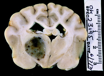
Signalment: 6-year-old female Boston terrier


History: Seven weeks before this examination, she had her first episode of generalized seizures, which occurred as a cluster over 2 hours. Clusters of seizures recurred at 10 days and 2 days before this examination, as well as in the hour before this examination. The owners described her as being normal between these clusters of seizures.


Examination: On the initial examination when she was presented to the hospital, she was depressed and blind, with normal pupil size and response to light. She drifted to either side as she walked and had slow postural reactions in all four limbs.


Anatomic Diagnosis: Diffuse brain but primarily the prosencephalon bilaterally


Because of the history of a seizure just before hospitalization and this examination, the concern was that these clinical signs represented the postictal effects of that seizure, and the examination should be repeated if and when she reached an interictal stage.


Second Examination: This examination was performed 24 hours after the initial examination. She was alert, responsive, and visual, and she had a normal gait. Hopping was consistently slow in the right limbs. Hypalgesia was found on the right side of the nasal septum.


Anatomic Diagnosis: Left prosencephalon, possibly the left frontoparietal lobe region that spared the left central visual pathway


Differential Diagnosis: Structural disease: neoplasm, necrotizing meningoencephalitis or leukoencephalitis, granulomatous meningoencephalitis, abscess, vascular compromise


Progressive seizures in a brachycephalic breed at this age suggest a neoplasm. Necrotizing encephalitides are uncommon in this breed. A focal form of the lymphoproliferative granulomatous meningoencephalitis is a possibility. A bacterial or fungal abscess or granuloma is uncommon. Be aware of brain infections with Coccidioides immitis in the southwestern states. Vascular compromise is unlikely to only cause a seizure at the onset and then cause progressive seizures.


Imaging procedures are necessary to define the clinical diagnosis further. Advanced imaging was not available at the time of this study. CSF contained 2 white blood cells (WBCs)/mm3 (normal <5) and 39 mg/dl of protein (normal <25), which supported a structural disorder and suggested that a neoplasm was more likely than an inflammatory process.


The patient was euthanized, and necropsy revealed a well-defined astrocytoma in the left side of the diencephalon at its junction with the telencephalon (Fig. 18-2).


Stay updated, free articles. Join our Telegram channel

Aug 26, 2016 | Posted by in INTERNAL MEDICINE | Comments Off on Seizure Disorders: Narcolepsy

Full access? Get Clinical Tree

Get Clinical Tree app for offline access