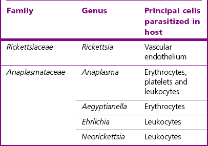
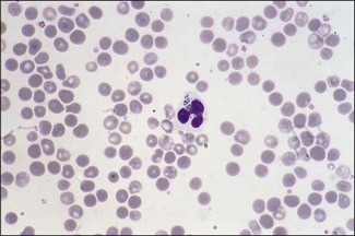
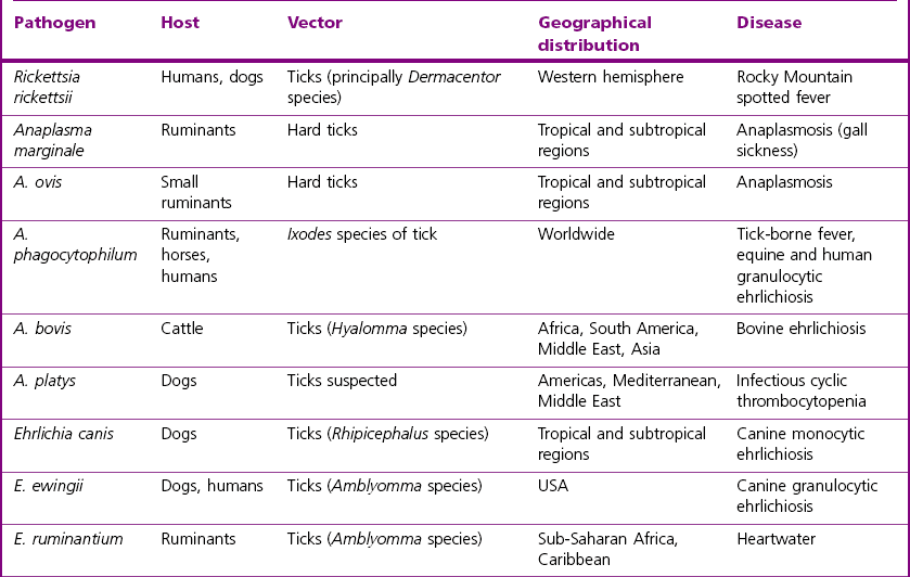
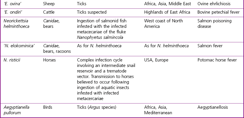
Chapter 34 Significant re-classification of the order has occurred in recent years (Dumler et al. 2001), much of it based on DNA sequencing information and in particular 16S and 23S rRNA gene sequence comparisons. The original tribe structure, the Rickettsiaceae family, has been eliminated, while members of the former tribe Ehrlichieae have been placed in the family Anaplasmataceae. The family Rickettsiaceae now comprises two genera, Rickettsia and Orienta while the family Anaplasmataceae comprises five genera; Anaplasma, Ehrlichia, Neorickettsia, Aegyptianella and Wolbachia. The status of the genus Aegyptianella is somewhat uncertain with recent data suggesting a close relationship to the genus Anaplasma. Several species have been re-assigned including Cowdria ruminantium to the genus Ehrlichia and Ehrlichia (Cytoecetes) phagocytophila to the genus Anaplasma. The genera Haemobartonella and Eperythrozoon, formerly assigned within the Anaplasmataceae, contain haemotrophic bacteria that lack a cell wall which have been shown to be closely aligned with species in the Mycoplasma pneumoniae group. These microorganisms now represent a new clade within the genus Mycoplasma (Neimark et al. 2001, 2002). The bacteria formerly known as HGE (human granulocytic ehrlichiosis) agent, Ehrlichia equi and Ehrlichia phagocytophila have been unified under the single species name of Anaplasma phagocytophilum. Coxiella burnetii, the cause of Q fever, is now known to be closely related to Legionella species and Francisella tularensis and has been re-classified to the gamma subgroup of the proteobacteria within the order Legionellales. For convenience, Coxiella burnetii is dealt with in this chapter as a separate section. Infection typically occurs as a result of the bite of an affected arthropod or following ingestion of infected flukes in the case of salmon poisoning and Potomac horse fever. The types of cells parasitized and disease pathogenesis varies with the rickettsial species (Table 34.1). Toxins, haemolysins and endotoxin-like lipopolysaccharides are known to be produced by some members of the group. Rickettsia rickettsii produces a phospholipase which aids escape from the phagosome soon after engulfment. Once in the cytosol nutrient acquisition and replication by binary fission is possible. The clinical signs of Rocky Mountain spotted fever are due to damage to the capillary endothelial cells, resulting in a necrotizing vasculitis and perivasculitis. Haemorrhages, thrombosis, oedema, loss of plasma and shock may follow. Ehrlichia species have a predilection for leukocytes and manage to replicate inside phagosomes by inhibiting fusion of the phagosome with a lysosome. When mammalian erythrocytes are parasitized, as in anaplasmosis or aegyptianellosis, anaemia probably results from the immune clearance of the altered erythrocytes. Anaplasma phagocytophilum and A. bovis are capable of surviving and multiplying within granulocytes while A. platys targets platelets. The principal diseases, hosts, vectors and geographical distribution of pathogens of veterinary importance in the Rickettsiales are summarized in Table 34.2. Table 34.1 Families and genera of the order Rickettsiales of veterinary importance and cell types targeted As members of the Rickettsiales stain poorly with the Gram procedure, Giemsa and other Romanowsky stains, such as Gimenez, Macchiavello and Leishman (Fig. 34.1), as well as fluorescent antibody (FA) staining (Fig. 34.2) are most widely used for both blood and tissue smears. A number of blood smears may have to be examined as the agents may only appear in the blood periodically. Figure 34.1 Leishman-stained blood smear from a sheep showing a neutrophil containing Anaplasma (Ehrlichia) phagocytophilum. (×1000)
Rickettsiales and Coxiella burnetii
Pathogenesis
Laboratory Diagnosis
Direct microscopy
![]()
Stay updated, free articles. Join our Telegram channel

Full access? Get Clinical Tree


Rickettsiales and Coxiella burnetii
Only gold members can continue reading. Log In or Register to continue
