4 Radiography
Radiography is a vital diagnostic tool in veterinary dentistry. Radiographs are required to:
DENTAL RADIOGRAPHY
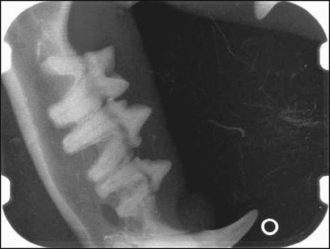
For a dental radiograph to be diagnostic, it should be an accurate representation of the size and shape of the tooth without superimposition of adjacent structures (Fig. 4.1). Intra-oral (film placed inside mouth and X-ray beam directed from outside the mouth through the tooth and adjacent structures onto the film) radiographic techniques are therefore required. The two basic techniques are:
Contralateral (same teeth, opposite side) views should routinely be taken for comparison.
EQUIPMENT AND MATERIALS FOR CONVENTIONAL INTRA-ORAL RADIOGRAPHY
The X-ray unit
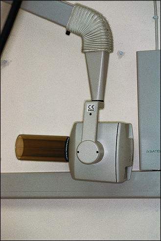
A dental X-ray machine (Fig. 4.2) is preferable to a veterinary X-ray machine. However, most veterinary X-ray machines can be used for dental radiography, but the film-focus distance will need to be adjusted to between 30 and 50 cm.
X-ray film
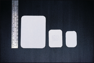
To allow intra-oral film placement and achieve high definition, dental film (Fig. 4.3) should be used. Dental film is single emulsion, non-screen, and is available in three sizes (occlusal, adult periapical and child periapical) and different speeds. The dental film is packed in either a paper or a plastic envelope, and the film is flanked by black paper and backed by a thin lead sheet (foil) that reduces scattered radiation.
Orientation
Each film has a raised dot in one corner. The dot helps with orientation when viewing and mounting dental radiographs if the following procedure is adhered to. Firstly, the dot should face the incident beam. Secondly, the film should be placed in the mouth so that the dot is always facing a specific direction. We position the dot so that it is always facing forward in the mouth. Another way of doing it is to ensure that the film is placed so that the dot is always in the same position, i.e. facing forward in the mouth on one side and backward in the mouth on the contralateral side.
Stay updated, free articles. Join our Telegram channel

Full access? Get Clinical Tree


