CHAPTER 21 Radiographic Considerations of the Young Patient
Radiographic Techniques
Progressive Puppy Skeletal Ossification and Growth
Figures 21-1 through 21-8 show the radiographic appearance of normal Cardigan Welsh Corgi puppies from a few days after birth to approximately 2 months of age.
3-Day-Old Puppy
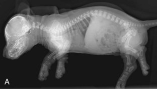
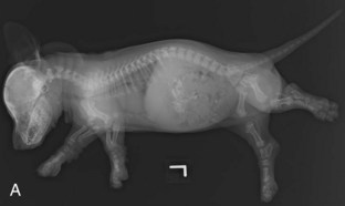
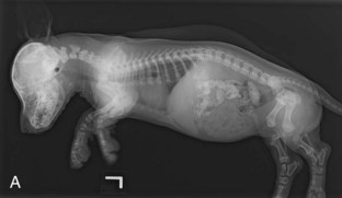
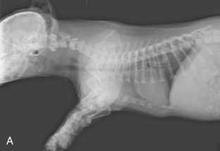
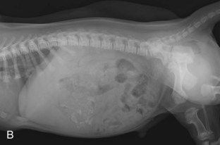
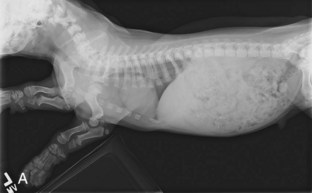
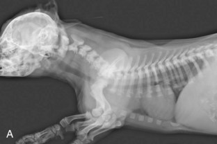
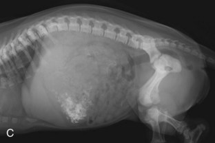
In a 3-day-old puppy (Figure 21-1), the heart looks relatively larger than in an adult (increased cardiothoracic ratio), as the lungs are not fully developed and well aerated as they will be later in life. As a result, the normal lung parenchyma shows a mild, diffuse, and unstructured interstitial pattern compared with an adult. Increased cranial thoracic soft tissue opacity is caused by overlying forelimb musculature and the thymus (see Figure 21-1, A). The abdomen of this puppy is normal and differs markedly from an adult. There is essentially no abdominal serosal detail, principally because of lack of abdominal fat to provide contrast with the soft tissue abdominal organs. Gastrointestinal gas provides the only contrast visible. The kidneys are not visible because of lack of retroperitoneal fat. The pendulous appearance of the abdomen is also a normal finding in puppies and kittens, caused by relative lack of abdominal wall muscle tone and a relatively large liver. In an adult, this appearance would be diagnostic for abdominal fluid.
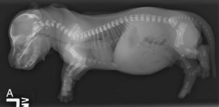
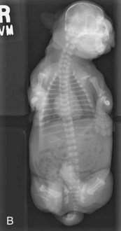
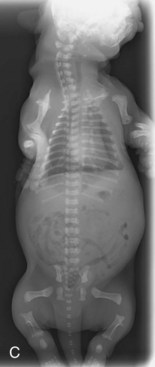
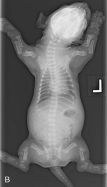
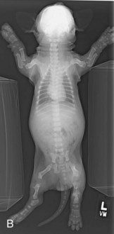
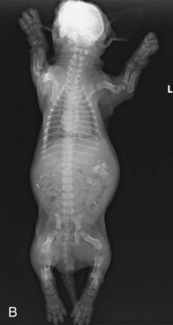
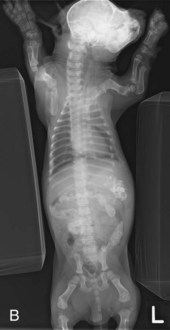
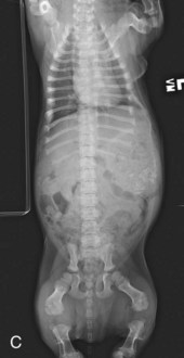
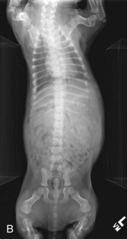
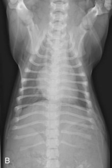
A VD whole body radiograph (see Figure 21-1, B) allows adequate visualization of the heart, lungs, and abdomen. There is increased width of the cranial mediastinum because of the presence of the immature thymus. When the puppy is repositioned to remove the overlying paws (see Figure 21-1, C), note the shortened, rectangular shape of the vertebral bodies created by nonossified vertebral endplates (epiphyses). The ilial and ischial ossification centers of the pelvis can be seen; the femoral heads are not ossified at this age.
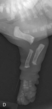
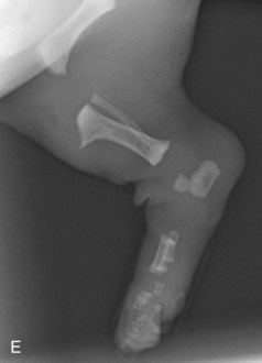
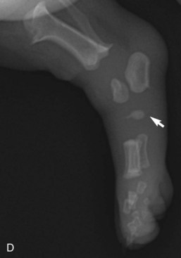
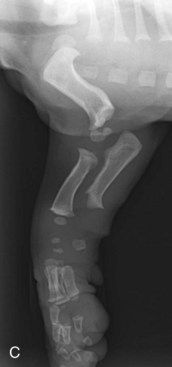
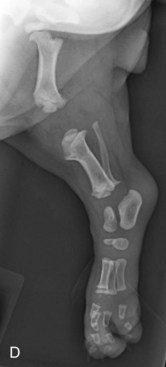
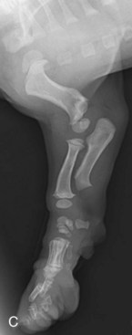
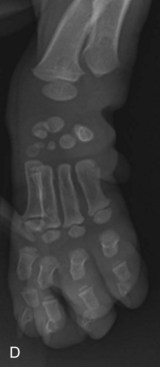
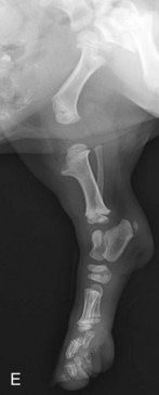
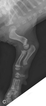
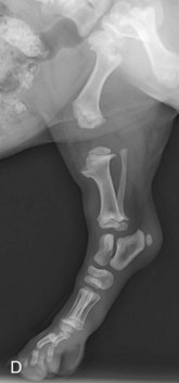
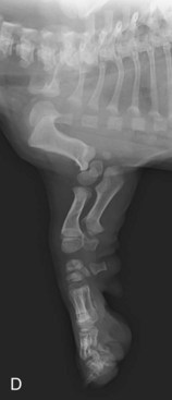
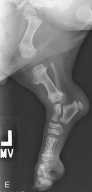
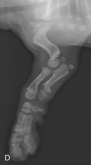
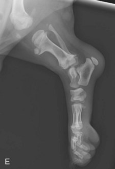
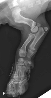
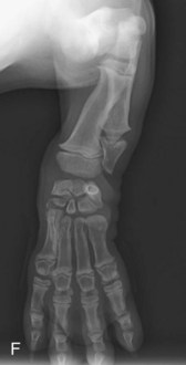
A lateral radiograph of the left forelimb (see Figure 21-1, D) shows that the humeral, radial, ulnar, metacarpal, and phalangeal diaphyses are ossified. The humeral head, distal humeral condyle (the lateral portion, the humeral capitulum, and the medial portion, the humeral trochlea, are separate centers, eventually fusing by approximately 2 months of age to become the humeral condyle), radial and ulnar epiphyses, metacarpal and phalangeal epiphyses, and all the carpal cuboidal bones are not ossified at this early age. Radiographs of the hindlimb (see Figure 21-1, E) show that the femoral, tibial, fibular, metatarsal, and first and second phalangeal diaphyses are ossified, as are the calcaneus and talus. The third phalanges are barely perceptible. All the epiphyseal centers are still cartilage models (soft tissue radiographic opacity) and not yet ossified. Note that the patellar, fabellar, and tarsal cuboidal bones are not visible as bony structures. In Figure 21-1, D and E, the lack of ossification creates a radiographic image of “widened” joints and soft tissue swelling. Therefore assessment for potential trauma or infection is challenging and may require comparison with the contralateral limb.
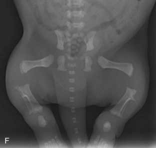
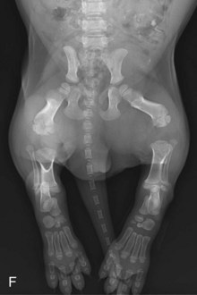
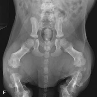
In a VD pelvic view (see Figure 21-1, F), the ilial, ischial, and pubic ossification centers are present, although the acetabular bones and femoral heads are not ossified. The stifle joints appear wide due to lack of epiphyseal ossification; the patellae and fabellae are also not ossified. The tarsi have a similar appearance, due to lack of epiphyseal and cuboidal bone ossification.
12-Day-Old Puppy
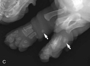
In the 12-day-old puppy (Figure 21-2), phalangeal and talus ossification has progressed. The primary difference in the radiographic appearance between the 3-day-old and 12-day-old puppy is identification of early ossification of accessory carpal bones (see Figure 21-2, C), the fourth tarsal bones (see Figure 21-2, D), and very faint ossification of the central tarsal bones. The cardiothoracic ratio is considered normal (identical to an adult) with better aeration of the lungs; although poor abdominal detail persists, a very thin stripe of abdominal fat can be seen separating the ventral abdominal body wall from the ventral liver margin, and the abdomen is less pendulous.

26-Day-Old Puppy
A lateral whole body radiograph of a 26-day-old puppy shows further skeletal ossification (Figure 21-3, A). The vertebral endplates (epiphyses) are now identified. Abdominal detail is still poor, but the thin layer of fat between the abdominal wall and the ventral margin of the liver is thicker and more evident than at day 12. In Figure 21-3, B, note visualization of carpal bones, many long bone epiphyses, vertebral endplates (epiphyses), and maturation of ossification centers noted 2 weeks earlier. The cranial mediastinum is widened by the thymus. Abdominal detail remains poor, but the margins of the spleen can be faintly seen along the cranial to mid-left abdominal wall. Partial ossification of the humeral head, distal humeral condyle (two parts), distal radial epiphysis, radiocarpal and distal row carpal bones, distal metacarpal epiphyses, and proximal epiphyses of the first phalanges has occurred in the forelimbs (Figure 21-3, C). The femoral head, distal femoral epiphysis, and proximal and distal tibial epiphyses are now clearly evident in the hindlimbs (Figure 21-3, D). The distal metatarsal and proximal first phalangeal epiphyses are faintly seen as tiny focal areas of ossification. Tarsal bone ossification has progressed.
34-Day-Old Puppy
In a lateral whole body radiograph of a 34-day-old puppy (Figure 21-4, A), the lungs are not fully inflated, resulting in a diffuse increased soft tissue interstitial pulmonary pattern, not to be confused with pneumonia. Cranial thoracic soft tissue opacity from the thymus and overlying forelimb musculature can be seen. Abdominal detail remains poor, but a small amount of intraabdominal fat is seen along the ventral body wall, separating several loops of small intestine. A VD whole body radiograph shows further skeletal ossification (Figure 21-4, B). Cranial mediastinal widening is caused by the presence of the thymus. Abdominal detail remains poor, similar to 1 week earlier. In the forelimbs (Figure 21-4, C), the humeral head is similar to what its mature shape will be. The distal medial and lateral portions of the humeral condyle (capitulum and trochlea) are readily identified as separate centers of ossification that will fuse to each other by approximately 2 months of age. The proximal radial epiphysis is beginning to ossify. Progressive ossification of the distal radial epiphysis, the carpal cuboidal bones, the metacarpus, and phalanges is noted. In the paws (Figure 21-4, D), continued maturation of the metacarpal bones and phalanges is evident, but the distal ulnar epiphysis is not yet ossified. Progressive ossification of the diaphyseal and epiphyseal long bones and the cuboidal bones of the tarsus has occurred in the hindlimbs (Figure 21-4, E). The apophysis of the calcaneus (calcaneal tuber) is now evident. Still not ossified are the patella, fabellae, and tibial tuberosity. A VD pelvic view (Figure 21-4, F) shows that the acetabular bones are not yet ossified, nor are greater trochanters, patella, and fabellae. The coxofemoral joints appear widened, as do all the joints imaged.
42-Day-Old Puppy
A lateral whole body radiograph of a 42-day-old puppy (Figure 21-5, A) shows how abdominal serosal detail continues to improve. Note the thin fat stripe between the ventral abdominal body wall and the ventral margin of the liver, as well as increased visualization of the serosal surfaces of the small intestinal loops, separated by mesenteric fat. The liver margin extends well beyond the costal arch, with smooth and sharply defined margins. Whereas in an adult this would indicate liver enlargement, this is a normal finding in puppies and kittens. Note the subcutaneous fat accumulation when compared with the earlier images.
In Figure 21-5, B, the thoracic portion of the puppy is mildly rotated with the sternum projected to the left. The thymus can be identified as a roughly triangular soft tissue opacity in the cranial left thorax. Abdominal detail continues to improve. Note the spleen is well visualized along the left lateral body wall by the thin fat opacity along its mesenteric margin.
Joints are becoming more adult-like in appearance as the epiphyseal bone continues to ossify (Figure 21-5, C). The distal ulnar epiphysis is now visualized; a tiny focus of ossification of the medial humeral epicondyle is now seen. The joints of the pes are becoming more adult-like in appearance (Figure 21-5, D). The stifle lags in ossification; the patella, fabellae, and tibial tuberosity have not yet begun to ossify.
49-Day-Old Puppy
In a 49-day-old puppy, progressive skeletal maturation has occurred. The thorax and abdomen (Figure 21-6, A to C) are otherwise very similar in appearance to the preceding week. In the forelimbs (Figure 21-6, D), the medial epicondylar apophysis of the distal humerus and distal ulnar epiphysis have significantly ossified during the past week. The remaining osseous structures are progressively maturing. Progressive ossification of the bony structures of the hindlimb has also occurred (Figure 21-6, E); however, the patella, fabellae, and tibial tuberosity are still not apparent radiographically.
55-Day-Old Puppy
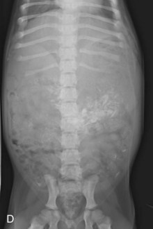
At 55 days of age, the thorax is normal (Figure 21-7, A). The vertebrae and other osseous structures continue to mature. A VD view (Figure 21-7, B) shows persistent widening of the cranial mediastinum caused by the thymus. Abdominal detail is still vastly different from an adult, but progressive fat deposition has resulted in a striking increase in abdominal serosal detail when compared with earlier images, although a typical puppy “pot belly” appearance remains (Figure 21-7, A and C). In the past 6 days, the olecranon (proximal ulnar apophysis) has been ossifying in the forelimbs. The supraglenoid tubercle ossification center is now visualized within the cranial portion of the scapulohumeral joint, overlying the manubrium (Figure 21-7, D). Within the same period, the patella and tibial tuberosity and the head of the fibula also now begin to ossify. Continued development of the remaining bony structures is evident. A VD pelvic view (Figure 21-7, F) will show that the acetabula continue to develop, although considerable ossification will occur during the next 2 months. The femoral heads are nearly completely ossified and have the shape of a mature dog. The patellae are radiographically evident.

62-Day-Old Puppy
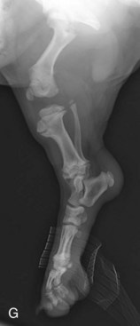
Once the puppy reaches 62 days of age, it is now large enough to justify a dedicated thoracic radiograph (Figure 21-8, A). The thorax resembles an adult in lateral views, with the exception of the increased opacity of the cranial mediastinum and the immature skeleton. The appearance of the intrathoracic structures closely resembles an adult dog on VD thoracic views (Figure 21-8, B) as well. The liver is sharply marginated and extends beyond the costal arch, a normal finding in a puppy or kitten (Figure 21-8, C). It is well delineated from the ventral abdominal body wall by an increasing layer of relatively radiolucent fat. Note that the serosal margins of various small intestinal loops are readily seen by surrounding mesenteric fat. There is not yet enough retroperitoneal fat to allow visualization of the kidneys, but there has been a progressive increase in subcutaneous fat layer along the puppy’s back. In a VD view, the serosal detail of the abdomen is not as apparent (Figure 21-8, D), and the kidneys cannot be seen. The supraglenoid tubercle of the scapula, the medial humeral epicondyle, the olecranon, and the distal ulnar epiphysis have enlarged and continued to ossify (Figure 21-8, E). The cuboidal bones appear very well ossified in a dorsopalmar view of the forelimbs (Figure 21-8, F). The paired palmar sesamoids of the metacarpophalangeal joints are now visible. The conical conformation of the distal ulnar physis is clearly apparent, as are the normal “flared” appearances of the distal radial and ulnar metaphyseal bone. Rapid maturation of the tibial tuberosity can be noted in the hindlimb (Figure 21-8, G), and the patella has developed a mature shape.
Stay updated, free articles. Join our Telegram channel

Full access? Get Clinical Tree


