8 Protozoal diseases
There are limited protozoal infections that affect the equine skin. These are invariably also geographically restricted. Whilst some such as besnoitosis and leishmaniasis have quite prominent skin implications, conditions such as dourine (trypanosomiasis due to T. equiperdum) have generalized clinical signs that are usually of far greater importance than the cutaneous signs. However, the cutaneous signs may be an early indication of some of the conditions and may also be the only outward sign of disease. Therefore the possibility of a protozoal diagnosis should not be forgotten.
Besnoitiosis (globidiosis/elephant skin disease)
Profile
This host-specific protozoal disease is due to either Besnoitia besnotii, or in donkeys B. bennetti, acquired by ingestion of feed contaminated with cat faeces, or by bites from blood-sucking insects and parenteral injection of blood from acute cases. Horses and donkeys have been reported affected in South Africa (Bigalke & Prozesky 1994) and North America (Terrel & Stookey 1973, Davis et al 1997).
1. Rare, geographically restricted skin disease of horses anddonkeys. Probably transmitted directly by biting insects.
2. Systemic signs including weight loss, depression and a nasal discharge may be present.
3. Multiple, small cutaneous papules with prominent flaking and scaling and more obvious papules in glabrous (hairless) skin.Some lesions have a glue-like exudate.
4. The organism in a biopsy of a topical papule is pathognomonic.
5. Potentiated sulphonamide may be effective when given for 30−60 days.
Clinical signs
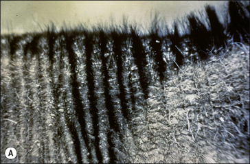
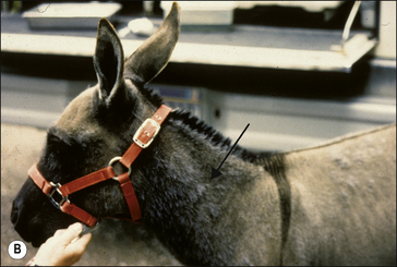
Chronic, pruritic, alopecic dermatitis with generalized thickening and lichenification of the ventral abdominal skin from sternum to scrotum or neck and withers (Fig. 8.1 and Fig. CD8 • 1) is characteristic of the disease. Multiple papules may be present in subcutis of perineum and abdominal area. The hair is sparse and the skin is inclined to crack and ooze. The worst lesions are usually around the fetlock. Small nodules may be present on sclera, soft palate, pharynx and larynx.
Exercise intolerance and bilateral nasal discharge may be present (van Heerden et al 1993). Other signs include an enlarged oedematous prepuce in males.
Differential diagnosis
• Dourine (Trypanosoma equiperdum): geographic restriction and venereal transmission are features; very easy identification of the parasites.
• Culicoides spp. hypersensitivity (sweet itch) and ventral biting Culicoides spp. hypersensitivity: sporadic and rare in donkeys, prominent seasonal management related pruritus.
• Equine viral arteritis: transient viral infection with minimal skin signs.
• Sarcoidosis: sporadic seborrhoeic disease with systemic signs. Diagnosis largely by exclusion of alternatives.
• Sarcoid (occult/verrucose forms): characteristic histology and typical appearance with multiple lesions over focal and sometimes more extensive areas in typical sites.
• Heavy metal poisoning: access and identifiable toxicological analysis in hair and hoof in particular.
Leishmaniasis
Profile
The causative organism is an obligate intracellular parasitic protozoan belonging to several members of the genus Leishmania including L. braziliensis, and is transmitted between horses by a few species of biting sandflies such as Lutzomyia cayennensis and Phlebotomus spp. The disease can, in theory at least, be transmitted to humans but there is little information available on this aspect (Yashida 1990).
In endemic areas such as South and Central America (particularly Brazil), southern Europe, and the Middle East, the cutaneous form of the disease occurs frequently in horses, donkeys and mules of all types and ages (van der Lugt & Stewart 1994). Isolated cases have been reported from the USA (Ramos-Vara et al 1996) and South Africa but it is extremely rare in non-endemic regions.
Stay updated, free articles. Join our Telegram channel

Full access? Get Clinical Tree


 Key points: Besnoitiosis


 .
. Key points: Leishmaniasis


