, Patricia A. Noguera1 and Trygve T. Poppe2
(1)
Marine Scotland Science, Aberdeen, UK
(2)
Norwegian School of Veterinary Science, Oslo, Norway
Abstract
Production related diseases and disorders cover a large area that the authors believe deserves increased attention, although for some areas we cannot cover the subject in great depth. These refer to a wide range of conditions that may or not be attributed to a biological agent with examples discussed in this chapter covering environmental related conditions, vaccination, developmental and congenital abnormalities and disorders, dietary imbalance, disorders affecting the heart and the eye, general skeletal abnormalities, and deformities among eggs and fry and predators.
Keywords
Production diseasesAbnormalitySalmonTroutProduction related diseases and disorders refer to a wide range of conditions with often multifactorial origin, and therefore they require a multidisciplinary approach in their management. Unspecific mortality varies from rather obscure causes like ‘loser syndrome’ or ‘failed smolt’ (Fig. 10.1), to other more specific causes such as poor smoltification, handling, transport, negative treatment effects and also certain infectious agents. A significant proportion of the mortalities can occur early post transfer to sea water and in some cases, might be linked to the quality of the smolt at the time of transfer, something that can and should be managed.
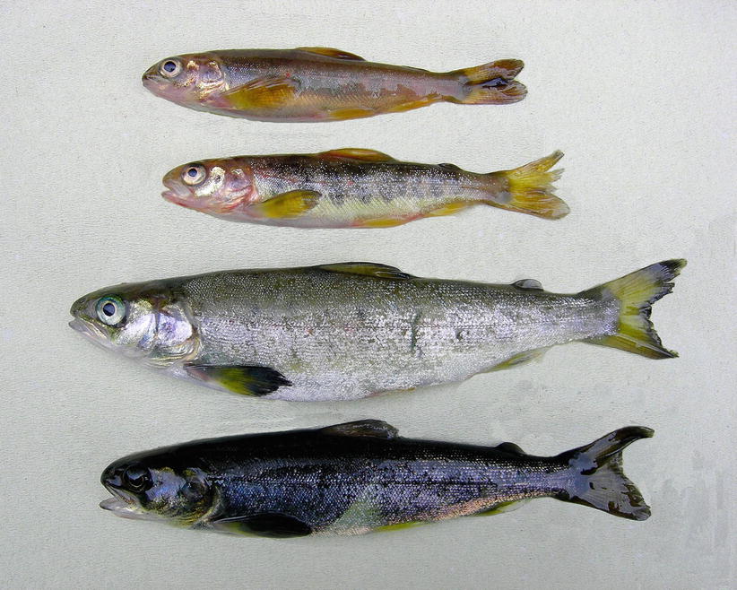
Fig. 10.1
Production Atlantic salmon ‘smolts’ recovered from well-boat during sea-water transfer
Overall, production related diseases and disorders cover a large area that the authors believe deserves increased attention, although for some areas we cannot cover the subject in depth. Examples discussed in this chapter cover environmental related conditions, vaccination, developmental and congenital abnormalities and disorders, dietary imbalance, disorders affecting the heart and the eye, general skeletal abnormalities, deformities among eggs and fry and predators.
10.1 Environment
10.1.1 Water Quality and Husbandry
Poor water quality is an obvious detrimental factor for any aquatic species including wild and farmed fish, with induced hypoxia being one of the major associated risks. Gill neuroepithelial cells (NECs) provide the sensory mechanisms for low oxygen detection, conversely, hyperoxia causes a decline in NECs number. These changes are concurrent with marked vascular distension, increased gill mucous, hyperplasia, elongation of respiratory lamellae and eventually, metabolic acidosis and death.
Fish excrete ammonia, and to a lesser amount, urea into the water as waste. Ammonia is highly toxic and exposure is a hazardous issue in fish farming resulting in a significant increase in oxygen consumption, with higher ventilation volume and respiratory distress. This can lead to acute mortality while the long term exposure results in reduced growth. Histologically, a severe branchial hyperplasia and associated widespread lamellar fusion, particularly at the tips, can be observed. The tissue may become oedematous, with a mild inflammatory response and occasional aneurysms.
In the farm environment, husbandry induced fin and skin damage can occur, although measures are generally in place such that these factors are kept to a minimum. Examples of production related damage in Atlantic salmon include frayed or damaged fins, scale loss and scrapes and are shown in Fig. 10.2
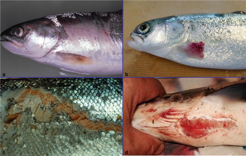
Fig. 10.2
(a) Net damage with scale loss in farmed Atlantic salmon. (b) Farmed Atlantic salmon with severe pectoral fin erosion with haemorrhage. (c) Farmed Atlantic salmon with skin wound after being scrapped on metal walkway. (d) Net scrape
10.1.2 Jellyfish
Fish reared in the marine environment can come into contact with true jellyfish. Depending on prevailing environmental conditions single animals or large swarms can be moved against the side of sea cages. Small jellyfish can be washed into cages whereas larger jellyfish tend to be break into pieces and tentacles or parts of tentacles enter the cages. In some cases the quantity of jellyfish can result in anoxia in the cages or obstruct respiration causing respiratory distress. Traumatic enucleation of the eye and marks on the side of the fish are also reported.
Several species of jellyfish have been associated with major loss of farmed Atlantic salmon, e.g. Aurelia aurita, Cyaneae capillata, Pelagia noctiluca and Phialella quadrata. Throughout European waters Muggiaea atlantica, P. noctiluca and Solmaris corona have contributed to significant loss in farmed fish. Blooms of C. capillata represent serious welfare issues due the irritant whiplash-like injuries inflicted by nematocysts of broken tentacles passing over the surface of the fish. P. quadrata (>15 mm in diameter) can pass through the mesh of sea cages and consequently sucked into the mouth of fish during respiration. At autopsy, as many as 40 jellyfish have been reported in the stomach of individual fish. In addition this species probably acts as a vector for the bacterium, Tenacibaculum maritimum .
Another species, Bolinopsis infundibulum has been reported to be associated with farmed fish mortality during the autumn in northern Norway. This species is fragile and ruptures on contact with the cages liberating a jelly-like substance which interferes with oxygen uptake by the fish gills.
Histologically, in general affected gills show sloughed lamella and necrosis, with oedema and inflammation of the filaments as emphasised by the presence of granulocytes. After approximately 48 h gill lesions show large areas of epithelial sloughing, haemorrhage and lysis of erythrocytes.
Cnidaria vary from a few millimetres to a few metres in size, and may be solitary (i.e. medusae of Hydrozoa, Scyphozoa and Cubozoa) or colonial (i.e. hydrozoan siphonophores) organisms.
10.1.3 Phytoplankton and Algal Blooms
Blooms occur naturally and can adversely affect human health as well as fish stocks. The blue-green algae, Cyanobacteria are among the most damaging blooms that impact on water quality. Impact depends upon the type, size and frequency of the blooms but reported as seasonal changes rather than influenced by fish farming. Fish stocks can suffer directly from toxins or indirectly through damage to the gill epithelia, resulting in acute necrosis, swelling, pyknosis and congestion.
10.1.4 Gas Bubble Disease
Gas bubble disease (GBD) is a non-infectious physically induced process that is caused by uncompensated hyperbaric pressure of total dissolved gases within the fish vascular system. GBD can either occur as a natural phenomenon in lakes and rivers (e.g. heating of water, photosynthesis) or artificially when supersaturated water is drawn into fish tanks without adequate aeration (e.g. pumping and heating of water, hydroelectric plants or leaking pumps). When pressure compensation is inadequate, a sudden decompression in the external environment (water) leads to blood dissolved gases (initially nitrogen) to form emboli in several tissues as it abruptly comes out of solution when trying to balance with the decreased external pressure. Highly vascularised tissues suffer most and severe exophthalmia due to the physical accumulation of gas bubbles in the choroid gland of the posterior uvea, is a frequent finding, as well as corneal degeneration and haemorrhage. Macroscopically bubbles may also be seen in the mucous membranes lining the oral cavity, the gills and fins (Fig. 10.3). Gas bubbles in the heart cavities can disrupt the blood flow and acutely affected fish subsequently die from asphyxiation. GBD can also lead to indirect problems and injuries when the air vesicles rupture (as in the skin and gills) leads to haemorrhage, open small wounds and secondary infections. Oedema of the lamellae with degeneration of the covering respiratory epithelium occurs with tissue necrosis and ischemia of the capillary beds. Safe limits for gas supersaturation depend upon the size of the fish, species, degree of super-saturation and water temperature.
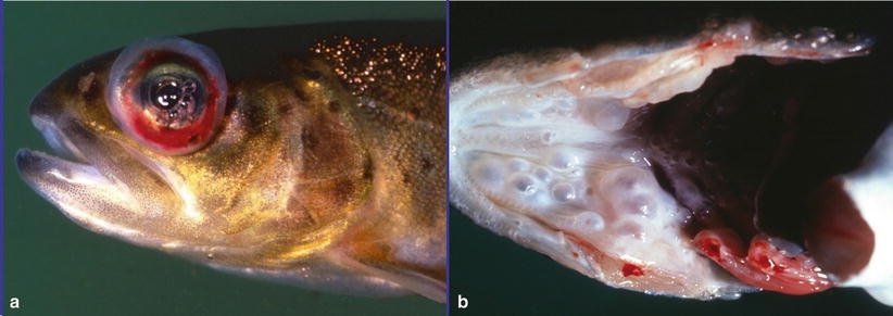
Fig. 10.3
(a) Exophthalmia, haemorrhage and ocular gas bubbles in brown trout with gas bubble disease. (b) Gas bubbles in the palate of brown trout with gas bubble disease
10.1.5 Nephrocalcinosis
Nephrocalcinosis occurs in intensively farmed salmonids, in particular rainbow trout and brook trout, but has also been seen in wild fish. The aetiology appears to be complex, but is often associated with high ambient free CO2 levels and/or nutritional aspects involving magnesium deficiency or selenium toxicity. Common signs in affected fish are abdominal swelling, exophthalmia and ventral haemorrhage which may continue to develop after transfer to sea water. At necropsy, ascites, splenomegaly and thickening of the ureters with white, chalky caseous deposits are frequent findings (Fig. 10.4). The kidney may become swollen, grey and urinary cysts can develop. Histologically, mineral deposits occur in distended distal tubules and ureters (Fig. 10.5). Deposits in adjacent parenchyma provoke a granulomatous inflammation with fibrosis and severe distortion of normal tissue. Mortality is generally low, but food conversion ratio in affected fish is impaired and the carcass quality is reduced. Diagnosis is based on gross lesions and the histopathological changes showing deposits in the collecting ducts which typically stain dark blue (basophilic) in H&E sections and black with von Kossa stain.
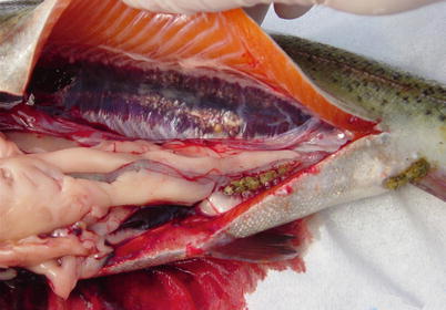
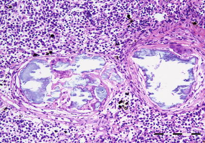
Fig. 10.4
Dilated ureters and granulomatous inflammation in the posterior kidney of farmed rainbow trout with nephrocalcinosis
Fig. 10.5
Nephrocalcinosis in farmed rainbow trout. Dilated ureters are filled with amorphous basophilic material. Bar = 100μm
10.2 Vaccination Side-Effects
With the exception of Tasmania, virtually all salt-water farmed salmonids are vaccinated. Most vaccines in current use are multivalent, i.e. they contain antigens from several different bacteria and/or viruses. In order to enhance and extend the duration and/or directing the nature of the immune response, an oil based adjuvant is mixed with the antigens. The composition of the vaccines may vary depending on the zoosanitary situation. After a few days of starvation fish are typically immunized prior to smoltification and sea-water transfer. The fish are anesthetized and the vaccine is given intraperitoneally, either by vaccination teams or automated machines. The injection site is typically in the ventral midline, 1.5 fin lengths cranial to the base of the pelvic fins, and the injection volume ranges from 0.05 to 0.1 ml.
The combination of antigens and adjuvant provokes a strong localized inflammatory response. This chronic inflammation is macroscopically visible as fibrinous strands between the peritoneal wall and internal organs (Fig. 10.6). Typically, the caudal parts of the pyloric caeca and spleen are involved in the lesions. Thrombosis and granulomatous inflammation is also reported in the liver. Moderate lesions are considered acceptable, both from an animal welfare and from the consumers’ points of view.
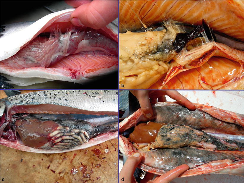
Fig. 10.6
Vaccinated Atlantic salmon showing. (a) Mild adhesions between visceral organs and body wall near the injection site. (b) Moderate adhesions between visceral organs and body wall; and (c–d). Severe melanisation of internal organs and peritoneal surface
Histologically, the affected region between the wall of the pyloric caeca and pancreatic tissue and around the spleen, show clear, empty spaces within the granulomatous response. These correspond to the oil droplets in the vaccine which are dissolved during the tissue processing. The inflammatory response is dominated by macrophages, lymphocytes, fibroblasts and multinucleated giant cells. Eosinophilic granular cells and melanomacrophages are usually present. In some cases, lesions become extensive and can involve most of the abdominal cavity with fusion of most organs to the body wall (Fig. 10.7). Extensive melanisation is sometimes evident. Granulomatous responses and location of oil droplets have also been recorded in organs such as gill, liver and muscle.
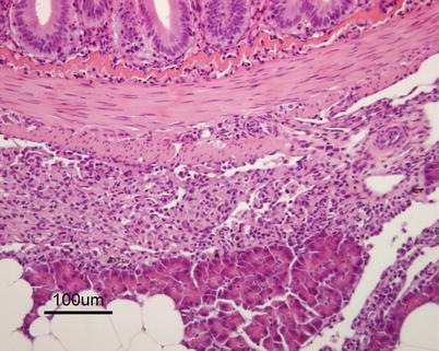
Fig. 10.7
Granulomatous peritonitis in Atlantic salmon vaccinated with oil-adjuvanted vaccine
Vaccination may also induce autoimmunity with marked glomerulonephritis (see Fig. 4.16) characterized by deposition of immune complexes in the mesangial cells of the glomerulus. Inflammatory responses may also be found in the uveal tract and in the heart (epicarditis and multifocal myocarditis). Occurrence of characteristic Splendore-Hoeppli reactions (asteroid bodies) in lesions is typical in hypersensitivity reactions.
10.3 Dietary Imbalance
Salmonids are, in principle, carnivores with a relatively short digestive tract and essentially identical, although slight modifications exist between species. Growth, food intake, stomach evacuation rate and feed efficiency ratio of Atlantic salmon are influenced by temperature and fish size. However, farmed and wild fish face very different challenges in relation to their nutrition. The former have a fairly continuous access to a dry pelleted feed throughout their life cycle and overfeeding results in overweight fish (Figs. 10.8 and 10.9). Conversely wild fish experience extreme variations in availability, quantity, quality and composition throughout the year and overweight wild fish are not observed. The composition and quality of pelleted feed is generally high, but scarcity of fish meal protein has resulted in their gradual replacement by plant components imposing a challenge to the feed manufacturers and to the fish. Quality control and customer demand have resulted in appropriate nutritional balanced diets for farmed salmonids and only rarely are deficiencies reported. Nevertheless, prolonged storage of feed under unfavourable conditions (e.g. heat, light, moisture) may compromise the quality and inconsistency may occasionally occur. In welfare terms there is also increasing evidence that intensively reared fish significantly alters some aspect of cardiac anatomy and physiology. In this section the effect of poor or deficient diets on the health of farmed fish is discussed.
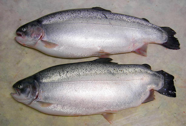
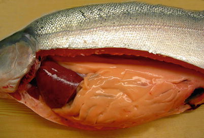
Fig. 10.8
Extremely obese seawater-farmed rainbow trout. The fish have frayed fins and the bottom fish an abnormality of the caudal part of the vertebral column
Fig. 10.9
Adipose tissue in the abdominal cavity of a sea-water farmed rainbow trout. Only the liver is visible; all other organs are hidden in fat
10.3.1 Inadequate Digestion of Pellet
Dry pellets will normally decompose to a porridge-like consistency in the stomach before passing to the pyloric region for further digestion, but at low temperatures, the feed is not always dissolved in the stomach and may become lodged as a hard plug in the sphincter area with resultant accumulation of dry and hard pellets in the stomach (Fig. 10.10). The mucosa is typically hyperaemic and irritated, which is also reflected in histopathological lesions.
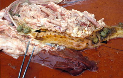
Fig. 10.10
Undigested pellets (arrow) in the anterior part of the rear intestine in farmed Atlantic salmon
Another condition that occurs under similar circumstances is where faeces of farmed salmon turn yellowish and foamy and float to the surface. The posterior gut is hyperaemic and the vent opening may be swollen. However, the direct causes of both conditions are unknown, but are believed to be associated to the technical and biological qualities of the pellet at variable ambient temperatures.
10.3.2 Hepatic Lipidosis
Farmed salmonids are susceptible to hepatic lipidosis or lipoid liver disease (LLD) when fed polyunsaturated fatty acids combined with insufficient amounts of antioxidant protection, such as vitamin E. Polyunsaturated fatty acids are prone to auto-oxidation on exposure to atmospheric oxygen.
Clinical signs include reduced appetite, impaired equilibrium and increased mortality. Fish become anaemic with pale gills and ascites, loose scales and develop fin necrosis. Internally, a yellow-orange discolouration and slight enlargement of the liver with a friable fatty consistency has been observed. The haematocrit is usually severely depressed as a result of a microcytic anaemia with increased blood cell fragility. Mortalities result from the cumulative effects of anaemia and hepatocellular and biliary tract dysfunction. Typical histological lesions show accumulation of excessive lipid in hepatocytes, macrophages and fat-storing cells (Fig. 10.11). Degenerating hepatocytes with pyknotic nuclei may occur over large areas. Red cell breakdown frequently results in haemosiderosis in the spleen. Diagnosis is based on internal appearance and evidence of a generalised fatty infiltration of the liver in stained tissue sections.
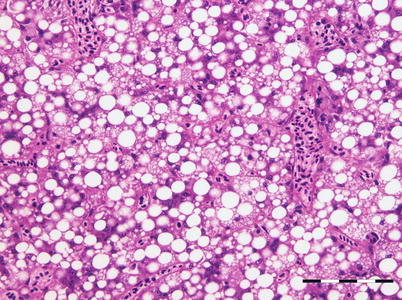
Fig. 10.11
Vacuolation of hepatocytes in farmed rainbow trout with fatty liver degeneration. Bar = 100μm
10.3.3 Steatitis
Steatitis or pansteatitis is an inflammation of the visceral adipose tissue that is normally abundant around the pyloric caeca in salmonids. The inflamed tissue is firm, yellowish or greyish and the swim bladder may be opaque with streaks of greyish yellow. Histological findings include thickening of the fat cell walls, infiltration, aggregation of ceroid-containing macrophages and pigment deposit (Fig. 10.12). A granulomatous response centred on multinucleate giant cells and eosinophilic inclusions may also be seen. Steatitis may occur as a sequel to pancreatitis, but also as an idiopathic disease where vitamin E deficiency and/or rancid fat in the diet are possible causes.
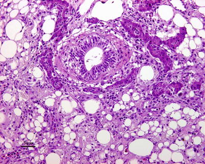
Fig. 10.12
Steatitis of peripancreatic fat in farmed Atlantic salmon
10.3.4 Heart Fat Infiltration
Fat infiltration in the epicardium can be encountered in both farmed and wild fish (Fig. 10.13). For example, wild whitefish may accumulate large amounts of fat, particularly between the ventricle and bulbus arteriosus and along the edges of the ventricle. In farmed salmon, the amount of epicardial fat is highly variable and is generally considered a negative trait.
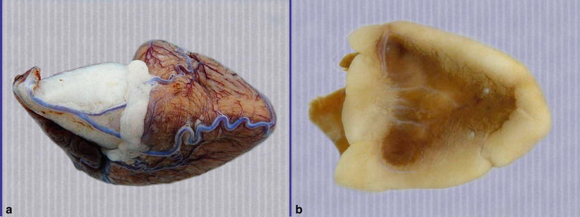
Fig. 10.13
(a) Congested coronary vessels and epicardial fat accumulation in the heart of farmed Atlantic salmon. (b) Extensive fat accumulation along the margins of the ventricle in wild whitefish (caudal view)
10.3.5 Soybean-Induced Enteritis
Salmon fed on diets rich in proteins from soybean can develop a non-infectious sub-acute enteritis affecting the distal intestine. Lesions include thickening of the lamina propria due to infiltration of macrophages, neutrophils, eosinophilic granular cells, loss of the normal supranuclear vacuolization of the absorptive cells in the intestinal epithelium and widening and reduction in height of the intestinal folds (Fig. 10.14). Absorption from the gut is reduced due to loss of apical vacuoles in epithelial cells. Lesions develop faster at 12 °C than at 8 °C and are generally resolved within a few weeks after removal of the soybean components from the diet.
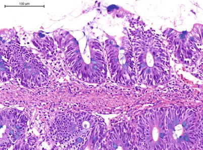
Fig. 10.14
Soybean enteritis in farmed Atlantic salmon
10.3.6 Vitamin Deficiencies
Ascorbic acid (vitamin C) is an essential dietary requirement which deficiency results in an impaired biosynthesis of collagen and chronic reduction of the supporting tissues, giving rise to a typical kyphosis, lordosis or scoliosis (Figs. 10.15 and 10.16). In some scorbutic fish, fractures in the axial skeleton with dislocations, focal haemorrhage and atrophy are noted (Fig. 10.17). A reduction in collagen and irregular hyalinisation of the remaining tissue results in a loose structure and dysplasia, including osteoporosis. Within the gill lamellae marked cell degeneration of the cartilaginous rod with large vacuoles accompanies deformation and distension (Fig. 10.18). Fish may show a hypoplastic anaemia and a decrease in haematopoietic tissue can be seen in the anterior kidney. Macroscopically, a shortened operculum is a common feature (Fig. 10.19). Gross appearance of the fish and the examination of histological sections provide provisional information for a diagnosis. An analysis of the vitamin content of the feed should be carried out to support a diagnosis.
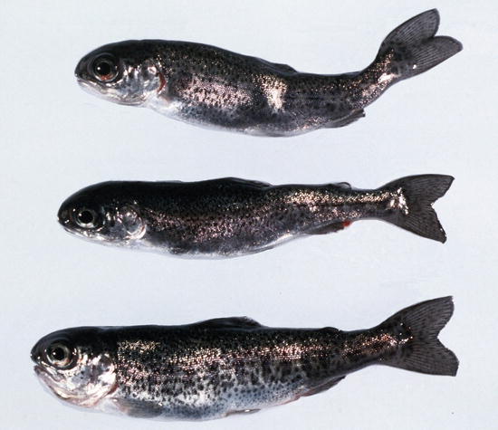
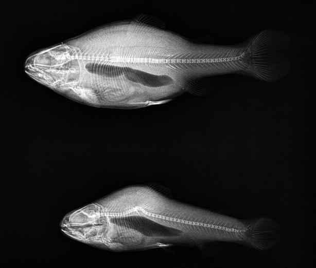
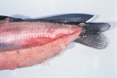
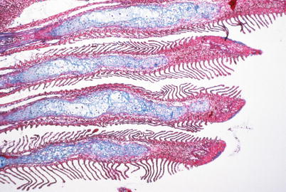
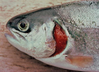
Fig. 10.15
Farmed rainbow trout fingerlings with vertebral abnormalities resulting from vitamin C deficiency
Fig. 10.16
X ray showing two different types of spinal deformity in farmed rainbow trout with vitamin C deficiency
Fig. 10.17
Abnormality of the caudal part of the vertebral column in farmed rainbow trout suffering from vitamin C deficiency
Fig. 10.18
Vitamin C deficiency in rainbow trout. The cartilage of the lamellae is curved and show irregularities. The cartilage cells are hydropic and vacuolated. Azan and toluidine blue. Low power
Fig. 10.19
Farmed rainbow trout with shortened operculum. Note exposed gill tissue
Vitamin E is an essential nutrient for many species of fish and deficiencies can occur where diets are partly oxidised before use. Several factors influence the dietary requirement for vitamin E, including the level of polyunsaturated fatty acids and other oxidants in the diet and fish tissues, as well as the level of selenium. A deficiency has an impact on several tissues including observations of white muscle degeneration (Fig. 10.20) and under experimental conditions, it is associated with calcification of the pseudobranch (Fig. 10.21).
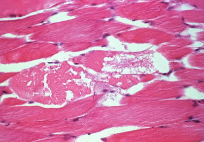
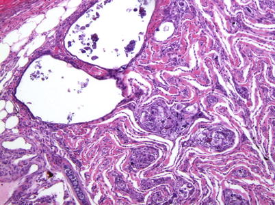
Fig. 10.20
Experimentally induced white muscle degeneration in Atlantic salmon with deficiency of vitamins C and E. Low power
Fig. 10.21
Experimentally induced calcification of the pseudobranch in Atlantic salmon with vitamin E deficiency. Low power
Pathological changes include lordosis, exophthalmia, splenomegaly, mottled liver, pale kidney, poor growth and ascites, and can be correlated with marked microcytic anaemia, reduced haematocrit and increased haemolysis. Incomplete maturation of these cells is consistent with increased fragility due the recognized role in cell membrane integrity and growth. In addition, steatitis has also been reported as well as small areas of degeneration with dilation of the liver sinusoids often with ceroid deposition. Splenic haemosiderosis is prevalent and occurs as a fine, stippled appearance to the tissue in Perl’s stained sections (see Fig. 4.22) and this correlates with the accelerated erythrocyte lysis. Vitamin E deficient salmonids are recognised from gross observations and the examination of stained tissue sections.
Histological abnormalities associated with pantothenic acid-deficient fish (vitamin B5) exhibit dermatitis and exudate-covered gill lamellae, with extensive hyperplasia (Fig. 10.22). Differential diagnosis include changes attributed to infestation with Paramoeba perurans (see Fig. 8.2).
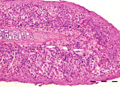
Fig. 10.22
Experimentally induced severe hyperplasia of gill epithelium and fusion of lamellae in rainbow trout with vitamin B5 deficiency. Bar = 100μm
Vitamin B6 deficiency in rainbow trout includes anorexia, listlessness, frantic erratic swimming, and ataxia. Deficiency can be readily determined by measuring pyridoxine-enhanced liver aspartate aminotransferase (ASAT) activity before clinical signs of deficiency are apparent. Histological changes include acinar cell atrophy (Fig. 10.23) and hyperplasia of renal haematopoietic tissue.
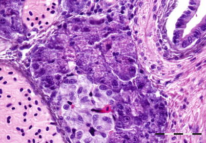
Fig. 10.23
Acinar cell atrophy in pancreas of Atlantic salmon with vitamin B6 deficiency. Bar = 50μm
Alack of thiamine (vitamin B1) is linked to the condition known as ‘M74’ in eyed eggs of wild Atlantic salmon and sea-trout in the Baltic area. The deficiency continues through the fry stages. Early mortality syndrome (EMS) and Cayuga syndrome are recognised as more or less identical conditions in several wild salmonids in the Great lakes area in North America. These conditions are characterized by up to 100 % mortality in progeny from certain female fish. Clinical signs include spiral swimming, loss of equilibrium and hyperexcitability, lethargy, dark body and subcutaneous oedema. Affected fry go off the feed and develop hydrocephalus, yolk-sac precipitate and haemorrhage (Fig. 10.24). Histologically, characteristic lesions are found in the molecular layer of the cerebellum developing cellular degeneration and necrosis, nuclear pyknosis and karyorrhexis, and sometimes haemorrhage. It is generally accepted that these lesions are the consequence of thiamine deficiency in the female. Salmonids are top predators and some of their prey may include forage fish with high levels of thiaminase, e.g. alewife in the Great Lakes region and sprat and herring in the Baltic Sea. The composition of the forage fish diet may be variable from 1 year to the other and individual fish may have different feeding strategies explaining the female dependant factor. High thiaminase levels results in low levels of thiamine in eggs and progeny with resultant characteristic signs of thiamine deficiency. These conditions can be prevented or reversed by exposing eggs or fry to thiamine. Females can also be injected with thiamine prior to stripping in order to avoid the condition. White spot disease and blue-sac disease are both differential diagnosis.
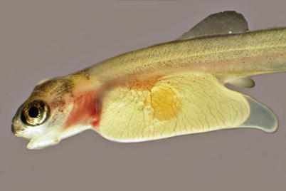
Fig. 10.24
M74 in Baltic salmon fry. Note haemorrhage in gill area
10.3.7 Mineral Deficiencies
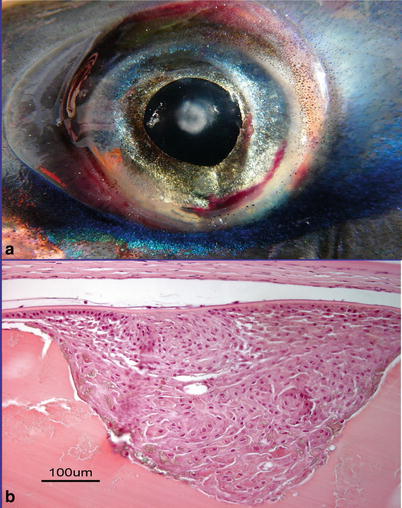
In aquatic ecosystems zinc is an essential micronutrient as well as a toxicant of considerable significance. Consequently, zinc deficiency leads to physiological perturbation of growth, reproduction, vision and immunity. Cataracts attributed to zinc deficiency are usually bilateral and characterised by diffuse or focal opacity of the lens. Other gross signs of this deficiency include a reduced growth rate with skin and fin erosion. Histologically, lesions consist of vacuolation, lysis of fibres and proliferation of capsular cells. Keratitis and panophthalmitis may sometimes occur concurrently. Cataracts occurring through zinc deficiency are recognised by a diffuse or focal opacity of the lens and the examination of stained sections. Diagnosis is based on histological assessment of eye sections.
Magnesium deficiency in rainbow trout include evidence of poor growth, loss of appetite, calcinosis of kidney and muscle and a dramatic increase in the muscle extracellular fluid volume. Histologically, there are degenerative changes in kidney, ovary and liver as well as hyperplasia of the renal haematopoietic tissue.
10.4 Diseases Associated with the Eye
A range of production related eye diseases are identified ranging from sub-acute lesions include cataract, anterior synechia and suppurative panophthalmitis (Figs. 10.25, 10.26, 10.27, 10.28 and 10.29). Lesions may be the result of haematogenous (endogenous) spread of pathogens, physical or mechanical (e.g. handling) with subsequent infection, chemical (e.g. medical treatments), thermal damage or nutritional factors.