Chapter 1 Principles of cell biology
Animal classification
If organisms have certain basic features in common they are grouped together into a kingdom. For example, if an organism is composed of more than one cell, i.e. it is multicellular, and obtains its food by ingestion, it is placed in the animal kingdom. Other kingdoms include plants and fungi. The animal kingdom is then further subdivided, based upon similarities of organisms, into a hierarchical system (Table 1.1). This narrows the classification down until we eventually reach a particular genus and species. Most living organisms are identified by a genus and species – a method known as the binomial system and invented by the Swedish scientist Carl Linnaeus.
Table 1.1 Classification of the domestic dog and cat
| Taxonomic group | Dog | Cat |
|---|---|---|
| Kingdom | Animal | Animal |
| Phylum | Chordata (Vertebrate) | Chordata (Vertebrate) |
| Class | Mammalia (Mammal) | Mammalia (Mammal) |
| Order | Carnivora | Carnivora |
| Family | Canidae | Felidae |
| Genus | Canis | Felis |
| Species | familiaris | catus |
| Common name | Domestic dog | Domestic cat |
These classes are then further divided into orders, and so on, until a species is identified, as in Table 1.1.
Generally speaking, all mammals have a similar basic structural plan in terms of anatomy and physiology, but each species has been modified to suit its specific lifestyle. In other words, mammals have become specialised for activities such as running, digging, gnawing, jumping and eating specific foods.
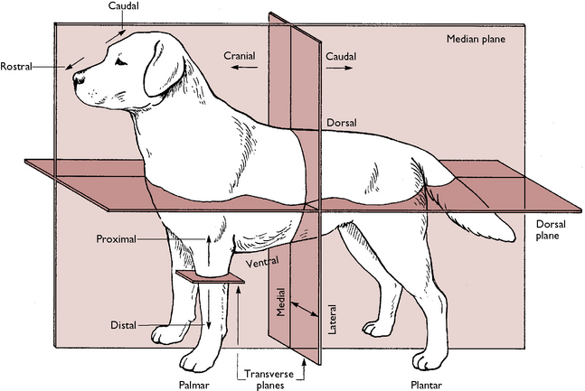
Anatomical definitions
When studying anatomy and physiology it is important to understand the terms that are used to describe where structures lie in relation to one another. These are illustrated in Figure 1.1 and named as follows:
The basic plan of the body
The body is made up of a number of systems and each of them has a specific job, enabling the body to function effectively. These systems can be placed in one of three groups depending on their function:
Structural systems
Coordinating systems
The mammalian cell
Cell structure and function
The components of a cell are shown in Figures 1.2 and 1.3 and are as follows:


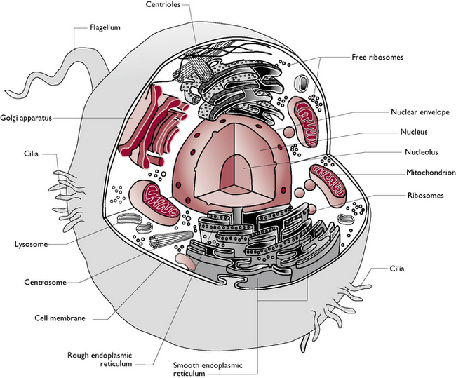
Fig. 1.2 Components of the mammalian cell.
(With permission from Colville T, Bassett JM 2001 Clinical anatomy and physiology for veterinary technicians. Mosby, St Louis, MO, p 11.)
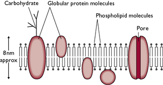
Cell membrane
The cell membrane covers the surface of the cell and may also be called the plasma membrane. It is responsible for separating the cell from its environment and controls the passage of substances in and out of the cell. Carbohydrates are found on the surface of the cell membrane and it is believed that these help cell recognition, e.g. they enable a cell to recognise whether or not it is in contact with another cell of the same type. The cell membrane of a mammalian cell is composed of a phospholipid bilayer (Fig. 1.4). This is a double layer of phospholipid molecules and has protein molecules embedded within it.
< div class='tao-gold-member'>
Stay updated, free articles. Join our Telegram channel

Full access? Get Clinical Tree


