CHAPTER 52Pregnancy Diagnosis in the Mare*
FERTILIZATION
The uterine tube has the ability to transport sperm toward the ovary while transporting the ovum in the opposite direction. Fertilization of the ovum occurs in the oviduct in mares. The fertilized egg or conceptus then takes 5 to 6 days to reach the uterus.
EMBRYONIC AND FETAL DEVELOPMENT
The conceptus is highly mobile throughout the entire uterus until day 16 after ovulation. The early movement of the conceptus is thought to play a key role in the physiologic event known as maternal recognition of pregnancy. The mobility phase ends when the conceptus becomes stationary and “fixation” occurs at the base of the right or left uterine horn. Fixation in postpartum mares tends to occur in the previously nongravid horn. Implantation occurs gradually and relatively late in the mare, beginning around day 40. It is not complete until approximately day 140. By convention, the term embryo is used up to day 40 and the term fetus after day 40.
Endometrial cups are a unique feature of the equine placenta. On or about day 28 of gestation, the chorionic girdle begins to form at the junction of the yolk sac and allantoic membranes. Specialized cells from the chorionic girdle invade the underlying uterine epithelium between days 36 and 38. Once in the endometrium, they enlarge and become clumped together to form the endometrial cups. The cups are arranged in a circle at the base of the gravid uterine horn. Endometrial cups produce equine chorionic gonadotropin (eCG), which stimulates the primary corpus luteum (CL) and causes induced secondary follicles to ovulate and/or luteinize. Because progesterone production remains high, mares do not show signs of estrus at the time of these secondary ovulations. This is independent from the presence of a fetus, so once the endometrial cups are formed, eCG production will proceed even in instances in which embryonic or fetal loss occurs.
ROUTINE PREGNANCY DIAGNOSIS
It is important to be able to tell if a mare is pregnant for management and husbandry reasons: Pregnant mares should be managed differently from nonpregnant mares. They should have further examinations to monitor the development of the pregnancy, confirm the absence of twin pregnancies, and monitor for early embryonic or fetal death. Twin pregnancies are bad news in the mare, and if the pregnancy is diagnosed early enough, appropriate intervention is possible (see Chapter 54).
It is just as important to identify the nonpregnant mare as soon as possible. Some mares have prolonged luteal activity (previously termed “persistent corpus luteum”), and the maintenance of circulating progesterone concentrations in these nonpregnant mares means that they do not spontaneously return to estrus. Persistence of luteal activity in the nonpregnant mare is a major cause of subfertility and is the main cause of anestrus during the breeding season. Traditionally, the term “prolonged diestrus” has been used to describe a condition in which the function of the CL continues beyond its normal cyclic lifespan of 15 to 16 days, resulting in the maintenance of elevated circulating progesterone concentrations for longer than expected. The term prolonged luteal activity is preferred, because “persistent diestrus” implies that the CL persists, whereas it is possible that other CLs are formed sequentially from diestrous ovulations. Prolonged luteal activity occurs in up to 20% of estrous cycles and are not accompanied by estrus; the cervix will remain pale in color, dry, and tightly closed. If diestrous ovulations occur late in the luteal phase, they will be refractory to the effect of endogenous luteolysins, resulting in a persistent luteal phase. Such abnormal luteal activity is associated with significant problems in reproductive management because affected mares may erroneously be assumed to be pregnant.
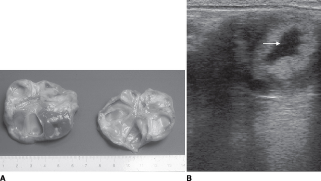
Plasma progesterone profiles are indistinguishable from those of pregnant animals. The uterus becomes firm and tubular (tonic) and the cervix is typical of that of pregnancy. Transrectal ultrasound imaging fails to detect a conceptus, but a CL can be detected in the ovary (Fig. 52-1, A and B). The ability to detect the CL throughout diestrus represents a profound diagnostic advantage of ultrasonography over rectal palpation.

Figure 52-1A, Gross specimen of left and right ovaries cut in half, showing a corpus luteum visible in the left ovary. A small central space can be seen in the corpus luteum. B, An ultrasound image of the equine corpus luteum with a centrally located anechoic area (arrow). The equine corpus luteum may remain uniformly echoic throughout its period of detection, or as in this image, an anechoic area may develop. In fact, a continuous distribution from uniformly echoic through to almost completely anechoic can be appreciated, with only a small border of luteal tissue.
Failure of synthesis and/or release of prostaglandin F2α(PGF2α) at the end of diestrus and failure of the corpus luteum to respond to PGF2αare the most likely causes of persistence of the CL. Treatment is by injection of a luteolytic dose of PGF2αor a synthetic analogue. The interval between treatment and ovulation varies considerably depending on the size of follicles at the time of treatment. Therefore, ultrasonographic examination of the mare before such treatment is always advisable in order to assess the status of folliculogenesis.
A clinical examination by a veterinary surgeon involving rectal palpation and ultrasound evaluation of the internal organs is the most accurate and useful method of pregnancy diagnosis. Nonetheless, several potential pitfalls have been recognized, including the confusion of uterine cysts for conceptuses and the presence of multiple conceptuses.
Speculum Examination of the Cervix
The vagina and cervix of the early pregnant mare are covered in pale, dry mucous membranes. The cervix is closed and resembles exactly the cervix of the mare in diestrus. As the pregnancy progresses, the external opening of the cervix becomes covered in tacky, dry mucus. Later in pregnancy (after 6 months), the cervix can be very relaxed.
Rectal Examination
Cervix
In early pregnancy (16 to 30 days), the cervix can be palpated on the floor of the pelvis as a rigid, firm structure (as during diestrus).
Ovaries
Both ovaries should be palpated, although ovarian palpation contributes little to pregnancy diagnosis. This is because both ovaries usually are enlarged from 18 to 40 days as a result of follicular development and the CL is not palpable. From 40 to 120 days, extensive ovarian activity with ovulations, luteinization, and development of secondary corpora lutea is evident. Follicular activity decreases from 120 days to term, and the ovaries become small and inactive. The position of the ovaries up to 60 days of pregnancy is as for the nonpregnant mare. From then on, they are drawn cranially and medially but remain dorsal to the uterus. From 5 months of pregnancy onward, the ovaries usually are not palpable.
Uterus
Both uterine horns and body should be palpated. Pregnancy diagnosis is based on tone and location of uterus and contents.
15 to 24 Days.
The uterus becomes more tubular and turgid from 15 to 24 days after ovulation and is readily palpable. It is not possible to feel the conceptual swelling. The uterus may be tonic in mares in a prolonged luteal phase and in nonpregnant mares bred at the foal heat, which are still undergoing uterine involution.
24 to 28 Days.
At about 26 days, the conceptual swelling develops at the uterine horn-body junction but can be difficult to palpate except in maiden mares. The swelling is 3 to 4 cm in diameter (about the size of a golf ball) and bulges ventrally.
28 to 35 Days.
The uterus is still turgid, and the conceptual bulge is more obvious. The uterine wall over the conceptus begins to feel thin.
35 to 60 Days.
As the conceptus grows, the swelling becomes larger and spherical and feels more like a fluid sac. By 42 days it is about 5 by 7 cm in size (the size of a tennis ball). The uterine tone around the bulge begins to decrease so that the swelling becomes less tense.
60 to 90 Days.
By 60 days, the conceptus swelling is approximately 12 cm in diameter and fills the pregnant horn. After 60 days, the pregnancy is like an elongated football and starts to involve the uterine body. The examiner must be careful not to confuse it with the bladder or a case of pyometra. The uterus migrates cranially.
Between days 60 and 100, the uterus is low within the abdomen, and the fetus usually cannot be palpated. From 4 to 5 months onward, the fetus usually can be palpated.
Ultrasound Examination
Rectal examination should always precede ultrasound examination. An initial rectal examination ensures removal of all fecal material, facilitates rapid location of the tract during scanning, and provides information on texture of structures.

Stay updated, free articles. Join our Telegram channel

Full access? Get Clinical Tree

