10 Pregnancy
She may have trouble whelping. Pups grow to fill the size available to them, and this pup had the whole uterus to himself. Singleton pups also may be associated with prolonged gestation because the single pup may not be able to set off the hormone cascade that initiates labor. Talk to your veterinarian about emergency care or performance of elective C-section.
There is no evidence that red raspberry tea leaves are beneficial during pregnancy.
I. PHYSIOLOGY AND ENDOCRINOLOGY
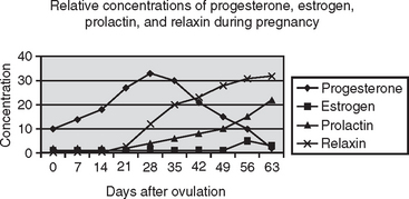
The eggs released by the bitch are fertilized in the uterine tubes and move into the uterus 8 to 9 days after ovulation. The conceptus implants and the placenta begin to be formed 16 to 18 days after ovulation. High concentrations of progesterone must be present throughout pregnancy. Progesterone during pregnancy decreases uterine contractility; stimulates secretions of the endometrial glands, which presumably provide nutrients to maintain the conceptus before implantation; and stimulates mammary development. Prolactin concentrations begin to rise at midgestation. Concentration of relaxin, released from the placenta, also rises beginning at midgestation (Figure 10-1).
Superfecundation is the presence in one litter of pups with different sires. This occurs readily in any bitch bred by more than one male. In some breeds, superfecundation is encouraged because it allows for greater genetic diversity in any given litter. This is especially useful in breeds with a small number of individuals. The American Kennel Club (AKC) does allow breeding a bitch to more than one male; genetic parentage testing of the pups is required before they can be registered (see Chapter 29).
Superfetation occurs when a pregnant animal is bred and a second conception occurs. This has not been documented to occur in dogs, nor is it likely because dogs ovulate all ova over a 24-hour period and because spermatozoa would be unable to reach the uterine tube past developed zonary placentas. If there are pups of grossly different size in one litter, it is most likely that the small pup had an abnormal placenta or developmental abnormality.
II. DIAGNOSIS
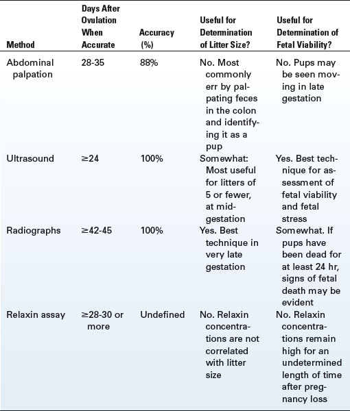
Pregnancy diagnosis in the dog is complicated by the fact that all dogs go through the same sequence of hormone changes after estrus, whether or not they are bred. For that reason, blood tests are not as commonly used to diagnose pregnancy as are diagnostic methods that physically identify pregnancy. The various methods of pregnancy diagnosis that are commonly used vary in time during gestation when they can be used, accuracy, and ability to provide information beyond a simple “yes or no” answer (Table 10-1).
Human “early pregnancy tests” do not work in dogs. These tests identify the hormone human chorionic gonadotropin (hCG), which is produced only during pregnancy in humans. The human conceptus implants 6 days after conception; so, by the time a woman suspects she may be pregnant, 14 days after ovulation, there has been a functional placenta secreting hCG for about 8 days. No hormone of this type is known to be produced in dogs during pregnancy. Because dogs do not develop a functional placenta until after implantation on about day 17 after ovulation, creation of an early pregnancy test for dogs is not likely.
A. ABDOMINAL PALPATION
Palpation of the abdomen permits the experienced individual to feel amniotic vesicles. The amniotic vesicles are the developing pups within the amniotic membranes, surrounded by fluid. These turgid vesicles have the consistency of peeled, hard-boiled eggs and range in size at midgestation from a grape in very small dogs to an egg in large-breed dogs. The vesicles are not grasped but instead are allowed to slip through the fingers as the abdominal contents pass through the fingers (Figure 10-2). Abdominal palpation never has been documented to cause pregnancy loss in bitches, but repeated palpation day after day is not recommended.
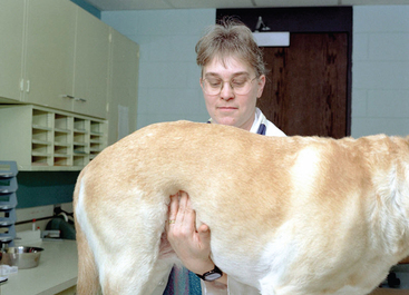
Abdominal palpation is best performed from about days 28 to 35 from ovulation. Amniotic vesicles are too small to feel much earlier than 28 days from ovulation. After about 35 days from ovulation, the amniotic vesicles fill with fluid and abut each other end to end, making them difficult to feel as individual entities. It is difficult to diagnose pregnancy in bitches that are tense, very thin, or very obese. Overall accuracy is reported to be 88%. If a mistake is made, it is more often a false-positive; feces in the colon can feel very much like amniotic vesicles, as can a segmented uterus affected with pyometra (see Chapter 16).
B. IMAGING
1. Ultrasound
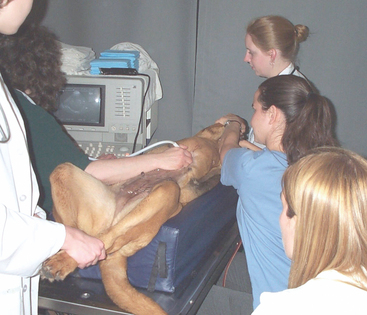
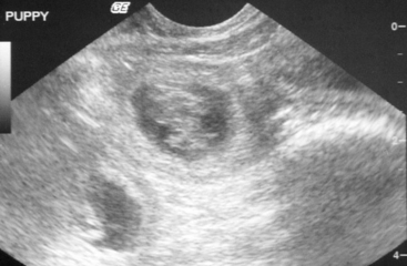
Several types of ultrasound are described in the veterinary literature. Those used commonly in small animal medicine are Doppler and B-mode ultrasound. With any type of ultrasound, sound waves are sent into tissue and reflected back. The reflection may be audible, as in Doppler ultrasound, or visible, as with B-mode ultrasound. Doppler ultrasound is rarely used as a diagnostic tool in veterinary medicine but is a component of systems designed for management of high risk pregnancies and possible dystocias (WhelpWise, Whelp Watch; see Chapter 12). For pregnancy diagnosis, B-mode ultrasound most commonly is used (Figure 10-3).
Amniotic vesicles easily are identified by B-mode ultrasound. The amniotic vesicles are balls filled with fluid, with a tiny developing puppy in the center. Fluid appears black on ultrasound, so B-mode ultrasound of a pregnant bitch’s uterus reveals multiple black spheres with gray material inside (Figure 10-4). There are reports of identification of pregnancy by B-mode ultrasound as early as 19 days after ovulation, but these amniotic vesicles are small and easily missed. Once the bitch is 24 days or more past ovulation, B-mode ultrasound is 94% to 98% accurate for pregnancy diagnosis.
Ultrasound is the best technique available for determination of fetal viability. B-mode ultrasound is “real time”; beating hearts and movement of limbs readily are identified after midgestation. Fetal heart rate also can be used as an indicator of fetal distress. Fetal heart rate should be about twice that of the dam, usually greater than 200 beats per minute. Fetal heart rates of less than 150 beats per minute signal severe fetal distress and the need for immediate veterinary intervention.
Stay updated, free articles. Join our Telegram channel

Full access? Get Clinical Tree


