7 Embryology
Embryology is the study of development from the fertilized egg to an individual. Some definitions are necessary to ensure understanding:
I. FERTILIZATION, IMPLANTATION, AND PLACENTATION
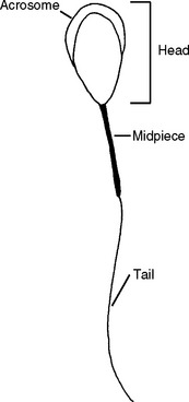
The spermatozoa introduced at breeding or artificial insemination are not capable of fertilizing the egg immediately on introduction to the female’s reproductive tract but first must undergo capacitation. During capacitation proteins are altered or removed from the head of the spermatozoon. Capacitated spermatozoa have enhanced motion, called hypermotility. The acrosome is a bag of enzymes covering the head of the spermatozoon (Figure 7-1). The acrosome reaction, which also occurs during capacitation, is dissolution of the membrane on the outer surface. This allows the enzymes to be exposed that will penetrate the outer layer of the egg during fertilization.
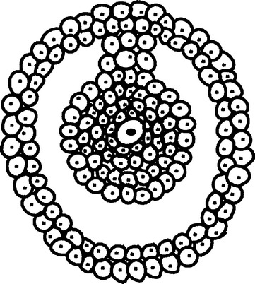
Capacitated spermatozoa reach the mature egg in the uterine tube. The ovulated egg is surrounded by a layer of cells from within the ovarian follicle. Within this layer of cells is a thick protein layer, the zona pellucida (Figure 7-2). The enzymes on the head of the spermatozoa and the hypermotile activity allow multiple spermatozoa to work their way through the outer cellular layer and approach the zona pellucida. As soon as one spermatozoon penetrates the zona pellucida completely and the inner membrane is reached, a reaction occurs that prevents any other spermatozoa from fertilizing the egg. The head of the spermatozoon that has penetrated the zona pellucida is drawn into the egg and its genetic material is released. Both the egg and spermatozoon contain half the genetic material of a dog. As the egg and spermatozoon fuse, the genetic material is combined and cell division begins.
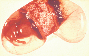
Placentas are classified by shape, amount of invasion into the uterine tissue, and amount of maternal tissue lost at the time of parturition (whelping). The shape of the canine placenta is zonary; the pup lies in a clear sac called the amnion, which is encircled with a strip of placental tissue that attaches like a ring inside the uterine horn (Figure 7-3). The pup is connected to the placenta by the umbilical cord. The margin of the placenta is a blood-rich band called the marginal hematoma, which is made up of stagnant pools of maternal blood that slowly leached into the space between the maternal and embryonic tissues. This may function as a source of iron for the developing puppies, but its exact function is unknown. The invasion type is endotheliochorial; the outer surface of the tissues arising from the developing embryo invades through the uterine lining and connective tissue to allow the bitch’s small blood vessels to grow directly into the placenta. The tissue-loss type is semideciduate; some maternal tissue is lost as the placentas detach at whelping.
< div class='tao-gold-member'>
Stay updated, free articles. Join our Telegram channel

Full access? Get Clinical Tree


