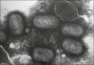
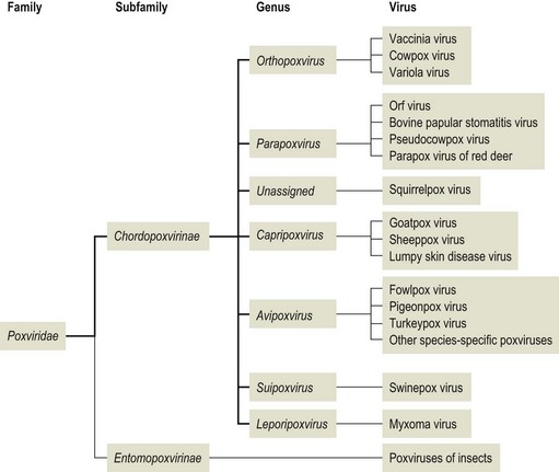
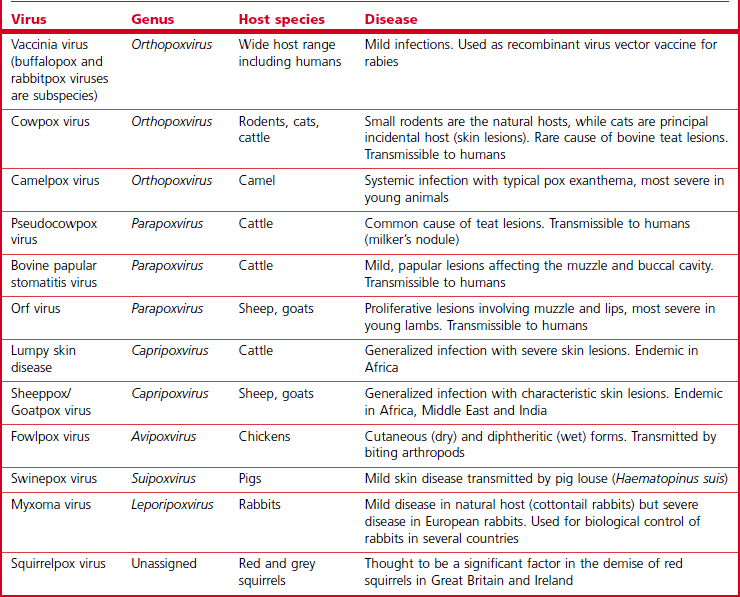
Chapter 51 The name pox is derived from an old English word ‘poc’ which means a vesicular skin lesion. Poxviral diseases typically affect the skin. Poxviruses are among the largest and most complex viruses with more than 100 proteins and several virus-specified enzymes. Members of this family infect many vertebrate and invertebrate hosts. Virions are large (220–450 nm × 140–260 nm) and usually brick-shaped with the external surface membrane containing lipid and displaying tubular or globular protein structures (Fig. 51.1); in contrast, parapoxviruses are ovoid with the surface membrane displaying a regular spiral filament (Fig. 51.2). Poxvirus symmetry is described as complex. There is a biconcave core or nucleoid that contains the linear, double-stranded DNA genome. One or two lateral bodies are present in the concave region between the core wall and the surface membrane. An additional cell-derived envelope may enclose the virus and these are termed extracellular enveloped virions (EEV). The family is divided into two subfamilies (Fig. 51.3), Chordopoxvirinae (poxviruses of vertebrates) and Entomopoxvirinae (poxviruses of insects). The subfamily Chordopoxvirinae is comprised of nine genera, Orthopoxvirus, Parapoxvirus, Avipoxvirus, Cervidpoxvirus, Capripoxvirus, Leporipoxvirus, Suipoxvirus, Molluscipoxvirus and Yatapoxvirus. Genetic recombination and extensive serological cross-reaction and cross-protection occurs within genera. Three closely related parapoxviruses, pseudocowpox, bovine papular stomatitis and orf virus, infect cattle, sheep and goats. These viruses are transmissible to man producing clinically indistinguishable lesions. The three viruses are morphologically identical and identification of the causal species relies on genetic studies. Capripoxviruses are economically important viruses producing generalized infections in domestic ruminants with significant mortality. The three members of the genus, sheeppox, goatpox and lumpy skin diesase virus, are closely related and share a group-specific structural protein named P32, making it possible to protect sheep, goats and cattle with a single vaccine. Squirrelpox virus was formerly assigned to the genus Parapoxvirus but has been removed and is presently unassigned. Poxviral infections are generally characterized by skin lesions (Table 51.1). Lesions may be localized to the teats and mouth or be multiple and distributed generally. Several virus-specified proteins are secreted from infected cells including a homologue of epidermal growth factor. This protein stimulates the proliferation of epidermal cells. Orthopoxviruses and swinepox virus produce lesions that begin as macules and progress through papules, vesicles and pustules to scabs that finally separate leaving a scar. The lesions produced by parapox, capripox and fowlpox viruses tend to be proliferative in nature with a granulomatous or nodular appearance. Infection with myxomatosis virus, a leporipoxvirus, is characterized by tumours. Cowpox virus is endemic in Europe. Although infection and disease have been described in cattle, cats, man and a range of captive mammals in zoological collections, these species are probably incidental hosts and the reservoir hosts are believed to be wild rodents (Chantrey et al. 1999). Genetic studies indicate that cowpox virus is probably a composite of up to five strains and support the creation of as many as four new species within the traditional ‘cowpox’ group (Carroll et al. 2011). Voles and woodmice are the principal reservoir hosts in western Europe. Clinical disease in cattle is rare and affects the teats of milking cows. Affected domestic cats usually come from rural areas and are described as good hunters. Infections in cats tend to peak in the Autumn when rodent populations are at their highest. There is often a history of a single bite-like wound on the head or a forelimb followed a few days or weeks later by widespread secondary skin lesions. Although cat-to-cat transmission can occur it is rare. These lesions begin as small papules but ulcerate over a period of two to three days. Scab formation follows with complete recovery usually in about six weeks. The diagnosis can be confirmed by histopathology, PCR, electron microscopy of unfixed scab or biopsy material and by virus isolation. Human infections with cowpox virus are uncommon and frequently associated with contact with infected cats. There is usually only a single lesion but affected individuals may be systemically ill. More severe disease and even fatal cases have been described in immunocompromised individuals. Bovine papular stomatitis is a mild viral infection of cattle that is transmissible to humans. Infection is believed to be common and worldwide. The virus typically produces a subclinical infection. Older animals are thought to serve as a reservoir of infection for successive generations of calves. Lesions are most commonly seen in calves on the mucous membranes of the buccal cavity and muzzle. They are characterized by hyperaemic foci that develop into papules with concentric zones of inflammation. Affected animals typically recover within three weeks. A more severe chronic form has been described occasionally and may be associated with concurrent infections or other immunosuppressive factors (Yeruham et al. 1994). The condition can be confirmed by electron microscopic detection of the typical parapoxvirus particles in lesion scrapings. The incubation period is about four to seven days. The virus is highly epitheliotropic producing proliferative wart-like lesions in affected animals following entry into the host through abrasions of the skin. The virus replicates in regenerating epidermal keratinocytes. Orf virus codes for a vascular endothelial growth factor which is thought to be important in stimulating vascular endothelial cell proliferation (Savory et al. 2000) and possibly providing the virus with further target cells to infect (Haig & Mercer 1998). Lesions progress through a series of characteristic phases. Initially papules develop but rapidly give way to a vesicular and then pustular stage. Scabs form within a few days while proliferation of the underlying dermis produces a verrucose mass. The lesions usually heal within four weeks leaving no scar. Secondary bacterial infection may prolong the course. The virus is readily transmissible to humans. A diagnosis of orf is normally possible on the basis of the characteristic clinical presentation. If necessary, electron microscopy can be performed on active scab material to confirm the diagnosis. Primers have been designed and used for the detection of parapoxvirus infections of ruminants by the polymerase chain reaction (Inoshima et al. 2000, Torfason & Guonadottir 2002, Gallina et al. 2006, Kottaridi et al. 2006).
Poxviridae
Cowpox virus
Bovine papular stomatitis virus
Orf virus
Pathogenesis
Diagnosis
![]()
Stay updated, free articles. Join our Telegram channel


Full access? Get Clinical Tree


