Postoperative Management

Chapter 7 Postoperative Management


Little has been written about proper postoperative nutritional management of the ruminant patient, even though all successful veterinary surgeons recognize its importance. In the absence of data from controlled clinical trials, this chapter attempts to record contemporary recommendations for postoperative feeding and nutritional support and is based largely upon the author’s opinions and experiences. Nevertheless, the recommendations made consider the physiology and pathophysiology of the relevant animal species and their clinical syndromes. The focus is on strategies for returning the gastrointestinal tract to normal function in the immediate postoperative period with less attention on specific long-term nutritional needs of the patient. More attention must be given to the nutritional needs of neonates and debilitated patients immediately after surgery, including partial and total parenteral nutrition. The nutritional support of these critical care patients is beyond the scope of this chapter.


Evaluation of Gastrointestinal Function


Evaluation of the gastrointestinal tract, especially the forestomachs and abomasum, is fundamental to postoperative evaluation of a ruminate patient. During the immediate postoperative period, the most important gastrointestinal tract features to monitor are gastrointestinal fill, motility, and microflora.


GASTROINTESTINAL FILL


In most surgical cases, the forestomachs are less full than normal because of anorexia or intentional preoperative fasting. Notable exceptions include vagal indigestion, carbohydrate engorgement, and obstructive diseases of the gastrointestinal tract. As appetite returns postoperatively, the rumenoreticulum and eventually the entire tract regain the appropriate fill. Gastrointestinal fill can best be evaluated by observing the abdominal contour facing the animal’s rear, combined with rectal palpation in cattle or transabdominal palpation and ultrasound examination in small ruminants. Extensive discussion of evaluation of abdominal distension can be found elsewhere. The most important abnormal contours of postoperative patients include the following: 1) dorsal and ventral distension on the left with ventral distension on the right; 2) prominent dorsal distension on the left with ventral distension on the right; and 3) bilateral ventral distension.


Prominent Dorsal Distension on the Left with Ventral Distension on the Right


Prominent dorsal distension on the left with ventral distension on the right usually indicates the same functional failures as the first abdominal contour, with the additional problem of free gas bloat or eructation failure. This can be confirmed by rectal palpation and passing an orogastric tube. Periruminal abscess and acites are two uncommon causes of this contour that must be differentiated from vagal indigestion. A periruminal abscess may occupy the left paralumbar fossa, thus giving the external appearance of bloat. Often, the diagnosis is missed or delayed because the history includes bloating in the past, which tends to lead the examiner to assume the distention continues to be caused by ruminal typany. There may also be a history of bloat treated with needle trocarization or intraruminal injection of antisurfactant products or by rumenotomy. Through the paralumbar fossa, the structure may even feel like a gas-filled rumen because of gas in the abscess. However, upon rectal examination, the examiner is unable to pass a hand between the rumen and left abdominal wall, and the abscess usually feels firmer than a rumen. Sonographic examination per rectum or transabdominally is useful, and centesis of the mass is confirmatory. In ascites secondary to uroperitoneum or peritonitis, the large amount of peritoneal fluid can push a rather small gas-filled rumen into the left paralumbar fossa and give the external appearance of rumen distension with bloat. Rectal palpation and abdominal sonography or centesis is confirmatory.


Bilateral Ventral Distention


Bilateral ventral distention usually indicates free abdominal fluid such as that accompanying uroperitoneum or diffuse peritonitis. It is similar to the second shape but without the dorsal distension and yields a more symmetrical shape. Sonography can differentiate abdominal fluid from intestinal ileus, another possible cause of this contour. Abdominocentesis will allow characterization of the fluid and definitive diagnosis.


The relationships between ruminal fill and feed intake and ruminal fill and fecal production are important to consider. If the rumenoreticulum fills rapidly with dehydration present or commensurate fecal production absent during the postoperative period, there is most likely a dysfunction of the forestomachs or abomasum. As mentioned previously, plasma electrolytes are often useful in assessing abomasal outflow, which may indicate abnormal abomasal motility, intestinal hypomotility, or obstruction. Other means of indirectly assessing abomasal and intestinal motility are auscultation, rectal palpation, and ultrasonography. The absence of gut sounds is a more reliable sign than the presence of sounds. The presence of sounds emanating from one area of the gastrointestinal tract does not mean that the other areas of the tract are moving, nor does it prove that the area producing sounds is functioning properly. However, the total absence of gut sounds can be a foreboding sign. Rectal palpation of the intestines does not detect motility, but fluid-filled intestines indicate functional or physical obstruction of the intestinal tract.


GASTROINTESTINAL MOTILITY


As defined in human medicine, postoperative ileus is abnormal motility of the gastrointestinal tract that follows almost every major surgical procedure, especially abdominal surgery. A postoperative paralytic ileus is a pathologic ileus that occurs after some surgical procedures and causes clinical signs and serious complications. In cattle, the rumen usually continues to contract during standing surgery; postoperative ruminal paralytic ileus is not usually a problem unless ruminal stasis was present prior to surgery. However, other parts of the ruminant GI tract are susceptible to ileus. No studies have been performed to define the duration of postoperative ileus in the different parts of the gastrointestinal tract of ruminants. In most species, the small intestine recovers within the first 24 hours and is followed closely by the stomach. The large intestine is the last region to recover. Understanding that gastrointestinal motility dysfunction always follows surgery and recognizing when expected “postoperative ileus” turns into pathologic and serious “postoperative paralytic ileus” is important.


Evaluation of gastrointestinal motility involves auscultation and palpation. Intestinal and abomasal sounds can be ausculted on the right side and ventrum, respectively. Rumenoreticular motility can be assessed by auscultation or by placing a fist in the left paralumbar fossa to feel the contractions. When evaluating ruminal contractions, one should note both the frequency and strength of contractions. The author prefers palpation to auscultation for initial assessment of ruminal motility. The normal rumen contracts about 2 to 3 times every 2 minutes. More complete evaluation of the rumen can be accomplished by combining auscultation and palpation through the left paralumbar fossa as well as rectally. In addition to frequency and strength of contractions, the physical character of the ruminal contents can be appreciated. A sutured surgical incision in the left paralumbar fossa, often present in postsurgical patients, may complicate the execution of auscultation and palpation in this area. The rumen has two contractile cycles that are called primary and secondary. The primary cycle is associated with mixing ingesta, while the secondary is associated with eructation. The primary cycle stratifies ingesta so the firm fibrous material floats in a mat on top of ruminal liquid. Small particles exit the rumen while larger ones are retained. Plant fibers more than 0.5 cm long in the feces indicate abnormal ruminal contractile activity. The primary cycle is under vagal parasympathetic control. Factors that stimulate contractions include feeding, low environmental temperature, and a slight distension sufficient to stimulate low threshold receptors in the rumen. The low threshold receptors can sometimes be exploited by pumping water and gruel into an empty rumen until mild distension is achieved. This helps stimulate ruminal contractions in anorectic ruminants. Factors that depress ruminal contractile activity include depression, fever, pain, endotoxin, volatile fatty acids, and abdominal distension sufficient to stimulate high threshold receptors. Ruminal motility can sometimes be improved by physically or pharmacologically reversing one or more of these inhibitory factors. Because these stimuli and suppressors of ruminal activity are mediated through the vagus nerve, an intact vagus is required for them to have an effect. Hypocalcemia reduces ruminal contractility by reducing the contractility of smooth muscle fibers irrespective of neural input.


Secondary ruminal contractions result in eructation of ruminal gas. They occur about every 2 minutes and are independent of the primary cycle contractions. Secondary contractions are stimulated by moderate distension and inhibited by severe distension. The secondary cycle can most easily be recognized by the occurrence of an eructation coincident with a contraction. Rumination is a specialized form of secondary contraction stimulated by coarse material in the reticulum and rumen. Rumination may actually be a source of pleasure for ruminants. Subjectively, the occurrence of rumination is a positive prognostic sign in the postoperative patient and is always a welcome sight in the eyes of this author.


The motility of the remainder of the ruminant gastrointestinal tract is similar to the nonruminant. The vagus nerve plays a significant role in controlling abomasal motility, but the intestinal tract is controlled principally by locally-produced substances and the enteric nervous system. In diseased animals, external factors such as electrolyte imbalances, inflammation, and endotoxemia can affect intestinal motility. In contrast to the simple and direct techniques for assessing ruminal motility, techniques for assessing abomasal and intestinal motility are indirect and not completely reliable. Auscultation, as described above, is perhaps most useful to confirm ileus through the absence of sound but is less reliable for confirming normal motility through the presence of sound. Ultrasound is being used in many species and holds promise for ruminants as well. Plasma electrolyte concentrations, especially chloride and bicarbonate ion, can be useful in determining functional or physical obstruction of the gastrointestinal tract. Severe hypochloridemia and alkalosis is usually associated with abomasal outflow problems, or obstruction or dysmotility of the orad small intestine. Less profound electrolyte abnormalities are observed in obstruction of the aborad small intestine and cecum.


RUMINAL MICROBES


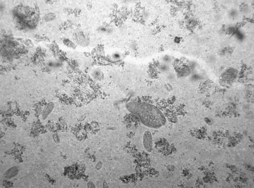
The ruminant forestomachs are a physiologic and biologic wonder, a marvelous example of symbiosis. In the mature cow, the rumen and reticulum represent 64% of the total stomach capacity, whereas the abomasum makes up only 11%. During the transition from preruminant to ruminant, the stomach changes in form, function, and fauna. The rumen and reticulum become a fermentation vat containing between 105 and 1012 bacteria/ml. The vast majority of ruminant microbes of animals on a forage diet are gram-negative anaerobic bacteria. The proportion of gram-positive organisms increases as the amount of grain in the diet increases. The ruminant’s ability to use poor quality roughage, inadequate to sustain nonruminant animals, is facilitated by the bacteria in the rumen. Although bacteria are more important for digestive function, the ruminal protozoa are easier to assess diagnostically, and they provide a reasonable index of ruminal health. Therefore a substantial proportion of the ruminal microbe examination focuses on the protozoal population. For clinical purposes, the ciliate protozoa can be divided into 2 morphologic types: holotrichs and entodiniomorphs. Holotrichs have cilia surrounding their one-celled bodies, whereas entodiniomorphs have cilia at one end (Figure 7.1-1).


Ruminal Fluid Analysis


Indications for clinical evaluation of ruminal microflora include suspicion of ruminal acidosis (e.g., carbohydrate engorgement), vagal indigestion, abomasal emptying defect of sheep, and rumen atony. Sometimes the analysis precedes surgery, but correction of the problem occurs during the postoperative period. A weighted stomach tube or a needle and syringe can be used to collect ruminal fluid for analysis. When a weighted collection tube is used, it is simply passed into the rumen, pushed back and forth to sink the tube, and aspirated. The first 100 ml or so is discarded to reduce salivary contamination. Aspirating transabdominally through the left flank by using a 16- to 18-gauge, 5-inch needle also helps eliminate salivary contamination.


A relatively simple analysis is sufficient for clinical evaluation of most presurgical and postsurgical cases. Color, odor, and smell should be evaluated immediately. Normal color is gray-green to green to brownish-yellow, depending on the diet. Milky gray or yellow fluid is associated with CHO engorgement. The pH of rumen fluid ranges from 5.5 to 7.0 in healthy cattle on a balanced ration. A pH paper with half-unit sensitivity is sufficient to diagnose ruminal acidosis or alkalosis of a single clinical case. A hand-held pH meter is required for adequate sensitivity at herd-level diagnosis of mild chronic acidosis. Cattle on high carbohydrate diets have lower pH values than those on roughage diets. Acid pH less than 5.5 in an anorectic ruminant indicates ruminal acidosis. Ruminal pH greater than 7.0 indicates ruminal alkalosis. Simple ruminal inactivity, or anorexia, results in ruminal alkalosis. Cattle with abomasal reflux may have an unusually low pH for an animal that has been off of feed for several days (e.g., 6.5 in comparison to an expected value of 8.0). This is because the abomasal acid has refluxed or been “vomited” into the rumen.


A very simple function test, the methylene blue reduction (MBR) time, can be performed rapidly without special equipment. The MRB test measures metabolic activity of the ruminal flora by indicating the relative redox potential of the rumen. One part of 0.03% methylene blue is added to 20 parts of strained ruminal fluid in a glass blood collection tube and is incubated at 37°C. A second tube of ruminal fluid serves as a control. Clearing of the dye in 5 to 6 minutes indicates active ruminal microbes. Delayed clearing indicates diminished anaerobic bacterial activity. In some cases, measurement of ruminal chloride is indicated. Ruminal chloride can be measured by standard electrolyte analyzers if the sample is filtered. It is elevated (>30 meq/L) in abomasal impaction and some other obstructive diseases in cattle and abomasal emptying defect in sheep.


Direct microscopic examination of fresh ruminal fluid on a slide is a quick and useful way to assess the health of the ruminal microflora. Abundant, live, active protozoa of various sizes and shapes will be present in cattle with a normal rumen (see Figure 7.1-1). Very large entodiniomorphs are the most fragile species; their presence suggests a healthy rumen. For further evaluation of the microflora, a drop of Lugol’s iodine can be added to a few drops of fresh rumen fluid. Lugol’s iodine kills the protozoa and stains carbohydrate in protozoa and bacteria. If the protozoa are depleted of carbohydrate, this indicates a depletion of carbohydrate in the rumen. Transfaunation of such an animal without concomitant force-feeding is likely to be ineffective because the newly introduced fauna will not have the substrate to allow them to multiply. Gram staining of the ruminal bacterial population, except in carbohydrate engorgement, has not been diagnostically useful in the author’s experience. If gram staining is performed on ruminal contents, one should expect to see primarily gram-negative organisms of a size and shape quite different from those encountered elsewhere in veterinary medicine. In CHO engorgement, chains of the gram-positive cocci Strep bovis proliferate first; then the large gram-positive rods of Lactobacillus sp become the predominant bacterial type.

Only gold members can continue reading. Log In or Register to continue

Stay updated, free articles. Join our Telegram channel

Sep 3, 2016 | Posted by in SUGERY, ORTHOPEDICS & ANESTHESIA | Comments Off on Postoperative Management

Full access? Get Clinical Tree

Get Clinical Tree app for offline access