CHAPTER 31 Postmortem Examination of the Puppy and Kitten
Why do a Postmortem Examination?
Puppy and kitten losses within the first few months of life are a common but unavoidable problem. Postmortem examinations provide essential information for preventative strategies and should be pursued aggressively. This is particularly important when losses occur in the preweaning period, and contributing factors should be eliminated before the next breeding cycle. Postmortem examinations are also an important part of quality assurance and quality control programs of veterinary clinics and provide great opportunities for continued education when faced with the unexpected or unexplained death of a patient or a patient’s death despite diagnosis-based treatment. Postmortem examinations address the owner’s concern about littermates and other pets in the household, the future of the breeding program, and the owner’s health. Results from a postmortem examination can also provide consolation, particularly after the difficult decision to have a pet euthanized.
What Comprises a Postmortem Examination?
A postmortem examination is actually a series of examinations. The gross examination during the necropsy of the animal is followed by the microscopic examination of fixed tissues (histopathology). Often additional tests on fresh tissues like microbiology (bacteriology, virology, parasitology, mycology, and serology) and/or toxicology (chemical analysis of tissues for toxins, minerals, and heavy metals) are necessary to determine the specific etiology of an identified disease process. These additional tests can be requested at time of necropsy, or samples may be collected and saved for testing at a later time, for example, when histopathology results are available and allow a focused approach to further testing to minimize expenses.
Who Performs Postmortem Examinations?
Ideally, the entire animal is submitted to a diagnostic laboratory for complete postmortem examination. If this is not possible, the necropsy may be performed at the clinic, and collected samples can be sent to a diagnostic laboratory for testing. Some of the advantages and disadvantages of the available approaches are listed in Table 31-1.
TABLE 31-1 Pros and cons of postmortem examination at a diagnostic laboratory versus at a clinic
Examination at | Advantages | Disadvantages |
---|---|---|
Diagnostic laboratory | Workup by specialists: gross lesions may be subtle and can be easily missed | Transport/shipping: expense and time in transit |
Coordination of ancillary testing | Additional cost (for gross examination) | |
Infrastructure for safe and complete examination | ||
Carcass: prearranged disposal or cremation | ||
Clinic | Immediate sample collection | Time consuming |
Immediate feedback | Biosafety and containment | |
Necropsy equipment and area | ||
Disposal of carcass; release to client not recommended | ||
Interpretation of findings (more often the challenge is the lack of significant gross lesions) |
Puppy and Kitten Losses and Common Causes
The pediatric patient poses some unique challenges with respect to postmortem examinations. The quick succession of developmental phases is associated with a rapid change of commonly encountered diseases to which the diagnostic approach has to be tailored. The perinatal puppy or kitten is commonly worked up as an abortion with a simplified gross examination and emphasis on histopathology and microbiological testing. In contrast, animals older than 1 day of age require a thorough and complete necropsy because gross findings determine the menu of additional tests to be performed. Before weaning, health concerns extend to littermates, the dam, and often the breeding program in general. After weaning, the problem is usually viewed as that of an individual animal and rarely of the household.
Very few studies on necropsy findings of the puppy and kitten have been published in the primary literature. In puppies, most deaths occur before rather than after weaning, which amount to more than 20% and less than 5% of all puppy deaths in the first year of life, respectively. More than 50% of the preweaning puppy losses occur during the first week of life. Common causes in this age group are stillbirth (incidence of 2.2% to 4.6%), trauma, failure to thrive, or congenital anomalies resulting in death or euthanasia. In puppies older than 1 week, respiratory and gastrointestinal diseases are the most common primary issues identified on postmortem examination, followed by malnutrition. In kittens, 50% of neonatal mortalities are the result of stillbirth (incidence of 4.3% to 10.1%), and an additional 25% occur in kittens less than 1 week of age. Approximately 15% of all litters have at least one kitten with one or more congenital anomalies (nearly 10% of kittens born alive). Based on histopathological examination, more than half of the kitten deaths up to 4 months of age are caused by infectious diseases, with the majority having a viral etiology. Before weaning, feline herpesvirus 1 and feline calicivirus are the main causes; after weaning, it is feline parvovirus followed by feline infectious peritonitis (corona)virus.
Box 31-1 highlights commonly encountered causes of canine and feline mortality by age group.
BOX 31-1 Common causes of mortality by age group
Abortion, Stillbirth, and Perinatal Phase (0 to 1 day)
Neonatal Phase (Puppies: 1 day to 10 days; Kittens: 1 day to 7 days)
Pediatric Phase


Figure 31-1 A, Moderate anasarca and severe hemoperitoneum, puppy. The subcutis is markedly expanded with edema (anasarca). The abdominal cavity is severely dilated with blood, most likely because of septicemia. Note how the umbilicus has been preserved in this partially skinned puppy. B, Cleft palate (palatoschisis), roof of oral cavity, dog fetus. C, Exencephaly, dorsal aspect of head and neck, dog fetus. The incomplete closure of the crania with protrusion of the brain through the defect in the bone and skin is often associated with cleft palate and other anomalies of the head. D, Bilateral hindlimb deformity and unilateral hydroureter, dog fetus. The abdominal cavity is partially exenterated, and the thoracic cavity is completely exenterated. The left ureter in the upper half of the picture is severely dilated with clear watery fluid (urine).

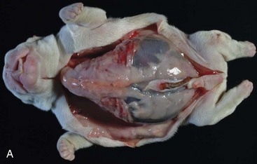
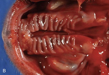
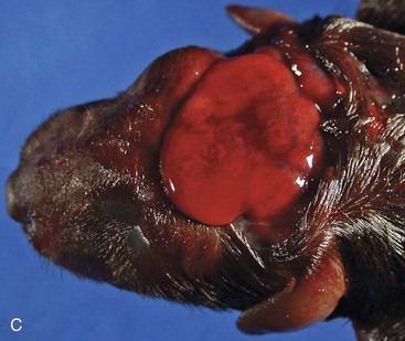
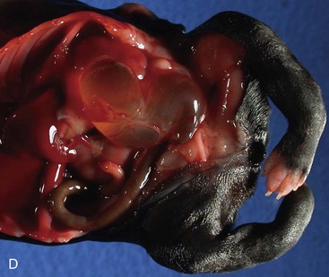
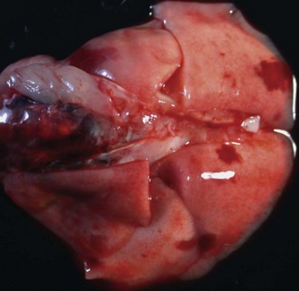
Figure 31-2 Multifocal hemorrhage, lungs, puppy. Compare these normally inflated and pink lungs with the pneumonic lungs depicted in Figures 31-3, B and 31-5. Cause of death in this puppy was septicemia.


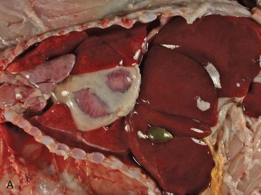
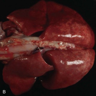
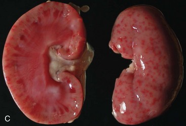
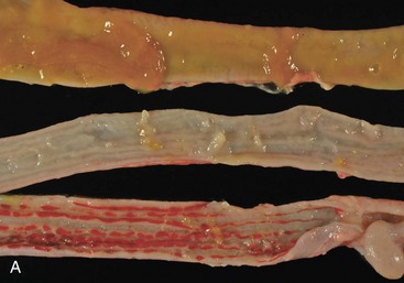
Figure 31-3 Systemic canine herpesvirus 1 infection. All the photographs in this figure show the same puppy. A, Severe acute hydrothorax and multifocal acute hepatic necrosis, situs of thoracic and cranial abdominal cavity. Pleural effusion and hepatic necrosis are common findings in herpesvirus infections. Canine herpesvirus infection is commonly diagnosed in puppies that died during the first 2 weeks of life. B, Severe acute interstitial pneumonia, lungs. The lungs are poorly collapsed and mottled red-beige. Compare with normally colored and collapsed lungs shown in Figure 31-2. C, Multifocal, severe, acute renal necrosis and hemorrhage, sagittal cut, kidney. Petechiation on the cut (left) and capsular (right) surface in the puppy is characteristic for an infection with canine herpesvirus 1.

Figure 31-4 Severe disseminated necrotizing hepatitis, abdominal situs, kitten. The liver is severely enlarged and mottled beige-brown. On histopathology, protozoan cysts consistent with Toxoplasma sp. were identified in areas of necrotizing hepatitis. The protozoal infection in this kitten involved many other organs, including the brain and lungs.

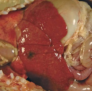
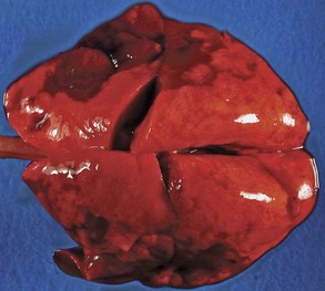
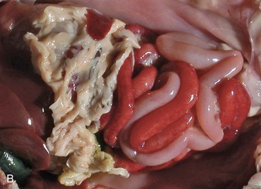
Figure 31-5 Severe subacute bronchopneumonia, lung, puppy. This gross presentation is typical for suppurative bronchopneumonia as a result of aerogenous infection with bacteria. Bordetella bronchiseptica was isolated in this case and is the most common bacterial pathogen in feline and canine respiratory disease. Compare with normally inflated and pink lung in Figure 31-2 and interstitial pneumonia depicted in Figure 31-3, B.


Figure 31-6 A, Necrotizing enteritis, jejunum and ileum, puppy. The Peyer’s patches in the affected flaccid segments of small intestine appear sunken because of loss of lymphoid tissue to necrosis. Parvovirus was identified in a mucosal scraping that included the malodorous intestinal contents. B, Segmental enteritis, abdominal situs, kitten. The severe hyperemia and dilation of loops of small intestine are typical for parvoviral enteritis (panleukopenia).
In a large number of cases, a definitive diagnosis is not established, and the cases are released as idiopathic deaths or abortions. In a retrospective study on kitten losses during the first 4 months of life, this apparent failure to identify a specific cause was significantly associated with submission of tissue samples instead of the entire animal to the diagnostic laboratory for postmortem diagnostics. This underlines the fact that a thorough gross examination and collection of a complete set of samples for histopathology and microbiology are imperative to successful postmortem diagnostics. Gross lesions may be subtle and can be easily missed by an inexperienced examiner. This may result in collection of incomplete sample sets, which in turn will limit the spectrum of available diagnostic tests.

Stay updated, free articles. Join our Telegram channel

Full access? Get Clinical Tree

