Stavros Yiannikouris, Claude A. Ragle
Postanesthetic Myelopathy
Postanesthetic myelopathy is an irreversible condition that leads to death or humane destruction of clinically affected horses. The condition, which follows general anesthesia, was first described in 1984 in a case report describing failure of spinal cord perfusion caused by a combination of hypotension and venous obstruction in the spinal cord. The suggested etiology was that the positioning during surgery resulted in pressure placed on the posterior vena cava by the abdominal viscera. Since then, occasional case reports have appeared in the literature, suggesting variations in the pathogenesis and proposing potential prevention and treatment protocols.
A recent survey of surgeons and anesthesiologists identified a number of cases and, together with the information presented in previously reported cases, described in greater detail the clinical signs, outcome, and histopathologic findings in horses with postanesthetic myelopathy.
Clinical Findings
There is no age- or breed-specific predilection for postanesthetic myelopathy, but large-framed or heavily muscled horses 2 years or younger are more commonly affected.
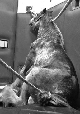
No preoperative abnormalities in the cardiovascular, respiratory, or neurologic systems have been identified in horses that went on to develop postanesthetic myelopathy in the postoperative period. Following general anesthesia in dorsal recumbency, affected horses most often develop flaccid limb paralysis. Horses show difficulty in rising or, in most cases, fail to stand. Affected horses usually have signs of pain and distress and make repeated attempts to rise. If they rise, they most often adopt a dog-sitting posture but are unable to adduct their hind limbs (Figure 209-1). A neurologic examination can differentiate postanesthetic myelopathy from myopathies or peripheral neuropathies by the absence of panniculus response from the midportion to the caudal portion of the thorax distally, by the loss of anal and tail tone, and by detection of areflexia and analgesia of the hind limbs. Incontinence and sweating, which extend from the midthorax caudally, can also be seen in a large percentage of affected horses. Neck and forelimb function are normal in most horses. Absence of the withdrawal reflex in the hind limbs, loss of deep pain, and absence of the panniculus response can distinguish postanesthetic myelopathy from bilateral femoral nerve paralysis, which may cause similar motor deficits.
Laboratory Findings
There are no preoperative signs with which to predict postanesthetic myelopathy, and no specific indices of subsequent disease have been identified in preoperative laboratory tests. Interestingly, in two horses in which serum vitamin E concentrations were measured, values were lower than reference range (2.5 to 3.5 µg/mL). In one of those horses, the blood selenium concentration was also slightly lower than reference values (0.10 to 0.25 ppm).
Postmortem Findings
Gross examination of the spinal cord usually reveals softening, discoloration, or both. Most lesions are seen in the caudal thoracic and lumbar segments, but some horses may have affected spinal segments in the cranial thoracic or caudal cervical spinal segments. Hemorrhage and congestion of vessels and spinal sinuses can also be seen. In some cases, no obvious gross changes are seen in the spinal cord. Only with histologic examination can a definitive diagnosis of postanesthetic myelopathy be reached. A histologic examination of spinal cord tissue should be performed in any horse that dies or is euthanized with a presumed diagnosis of postanesthetic myelopathy.
In most cases, histologic examination shows asymmetric or symmetric poliomyelomalacia, which can vary in severity from mild degeneration to severe necrosis of neurons, mostly in the ventral gray matter. Detection of hemorrhage and congestion of vessels in the spinal cord gray matter is less consistent. The white matter of the spinal cord is usually spared, but in some cases, edema and swelling of the axons can be identified. Lesions are located in the caudal thoracic and lumbar segments of the spinal cord, but more cranial segments can also be affected.
In the small number of horses in which the nucleus cuneatus of the brainstem was examined, degenerative neurons and spheroids were observed. Lesions in this location may implicate a causative role for vitamin E deficiency.
Traumatic injury of the spinal cord, such as occurs in postanesthetic myelopathy, can cause stretching and tearing of blood vessels, which results in myelomalacia and hemorrhage in the tissues. Because this lesion can develop 12 to 24 hours after injury and may progress both cranially and caudally from the original site of trauma, the length of time that elapses between the end of the anesthetic period and death affects the histologic changes that are observed postmortem. Acute malacic lesions, in the absence of hemorrhage, may not be histologically demonstrable if this time is less than 12 hours.


