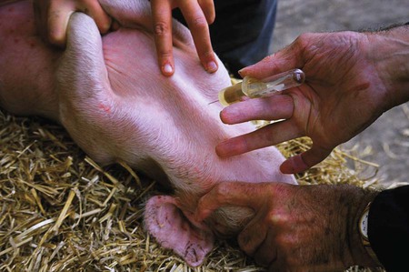
When you have finished this chapter, you will be able to • Set up and prepare the patient for each procedure, perform the procedure (when appropriate), or assist the clinician in performing diagnostic sampling and medication procedures • Successfully and safely administer medications by oral, nasal, and parenteral routes • Properly insert and maintain an intravenous catheter and monitor the catheter for complications • Explain the rationale and indications for each of the clinical procedures described • Set up materials and equipment and prepare the patient as needed for the procedure • Provide assistance to the veterinarian with performing the procedure or perform the procedure when it may be appropriate for a veterinary technician to do so • Perform or assist with necropsy and sample collection procedures and maintain a safe environment during these procedures The vein runs near the lateral border of the ear pinna and is accessed from the dorsal (haired) side of the pinna (Figs. 23-1 and 23-2). It is easily visualized and can be distended with finger pressure at the base of the lateral surface of the ear, although both hands of the technician can be freed up by using a mechanical method to distend the vein. A rubber band can be placed as a tourniquet around the base of the ear to distend the vein. Another method uses self-retaining forceps with long, plastic-covered jaws that are placed across the base of the ear to distend the ear vein. This method leaves one hand free to stabilize the tip of the ear while the other hand places the needle and aspirates the blood. The needle should enter the vein at a 45-degree angle to the skin. The ear vein may continue to bleed for several minutes after venipuncture is completed. Larger animals are restrained while they are standing, usually with a hog snare. The head should be raised slightly. Alternatively, some farms use a bleeding chute with a head catch. Nonslip footing should be provided. If the pig sits down before or during the procedure, stop the procedure, withdraw the needle, and get the pig on all four feet. Sitting alters the anatomical landmarks for the procedure and makes it difficult to perform successfully. An 18- to 20-ga × The venous sinus is located adjacent to the medial canthus of the eye and can be used for venous blood collections in pigs of any age. Approximately 5 to 10 ml of blood can be obtained. Small pigs are restrained in dorsal recumbency, inclined with the head down, and firmly restrained. Larger pigs are restrained standing with a hog snare. Piglets require a 20- to 20-ga × 1-inch needle. Larger pigs require a 16- to 20-ga × Normal complete blood count and blood chemistry values for swine are listed in Tables 23-1 and 23-2, respectively. TABLE 23-1 Complete Blood Count Normal Values for Swine
Porcine Clinical Procedures
Diagnostic Sampling
Venous Blood Sampling
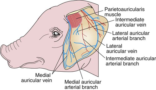
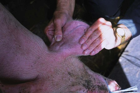
Lateral Auricular Vein
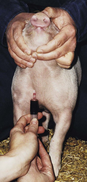
Cranial Vena Cava
 -inch needle is used for feeder (finisher) pigs weighing more than 50 lb. In adult swine, a 16- to 17-ga × 4- to
  -inch needle is needed. The syringe is attached for the procedure. The technician kneels in front of the animal on the right side (facing the body of the pig) or to the side of the right shoulder (facing the neck). The needle is inserted and directed exactly as described earlier (Figs. 23-3 and 23-4). Once the skin has been penetrated, slight backpressure is maintained on the syringe plunger. Blood flows readily when the vein is entered. In adult pigs the vein lies quite deep, up to 4 inches.
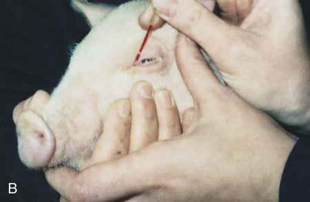
Orbital Sinus (Medial Canthus of the Eye)
 -inch needle. The needle is inserted deep to the nictitating membrane (third eyelid) and advanced at a 45-degree angle toward the opposite jaw. The needle will hit the lacrimal bone. Rotate and slightly withdraw the needle until blood flows from the hub. The needle is then attached. Aspiration should be gentle (Fig. 23-5).
Cephalic Vein
Packed cell volume (%)
32–50 (42)
Hemoglobin (g/dl)
10–16 (13)
Red blood cells (× 106 /µl)
5–8 (6.5)
Total protein (g/dl)
5–8 (6.5)
White blood cells (× 106 /µl)
11–22 (16)
Platelets (× 106 /µl)
3.25–7.15 (5.2)
Mean corpuscular volume (fl)
50–68 (63)
Mean corpuscular hemoglobin (pg)
16.6–22
Mean corpuscular hemoglobin concentration (g/gl)
30–34 (32)
Bone marrow (myeloid to erythroid ratio)
1.77 ± 0.52 : 1
DIFFERENTIAL, ABSOLUTE
Segs
28%–47%
3000–10,500
Bands
0%–2%
Lymphocytes
39%–62%
4300–13,700
Monocytes
2%–30%
220–2200
Eosinophils
0.5%–11%
0–2500
Basophils
0%–2%
0–400![]()
Stay updated, free articles. Join our Telegram channel

Full access? Get Clinical Tree


 -inch needle can be used. Small pigs up to 50 lb require an 18- to 20-ga × 1- to
  -inch needle. Small pigs are placed in dorsal recumbency with the head held firmly still. The front legs are extended and pulled caudally for complete access to the caudal neck and shoulder area. The needle is inserted (syringe attached) on the right side, at the caudal extent of the right jugular furrow, just lateral to the manubrium of the sternum. The needle is directed toward the caudal aspect of the top of the opposite (left) shoulder blade. Slight backpressure (vacuum) is kept on the syringe. The vena cava is encountered at a depth between
  and 2 inches, depending on the size of the animal. Blood is easily aspirated when the needle enters the vein.
 inches in piglets to 16 ga × 3 to
  inches in mature pigs.
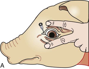
 -inch needle is sufficient. Up to 10 ml of blood can be obtained.