CHAPTER 12 Pigmentary Abnormalities
Terminology and introduction
The color of normal skin depends primarily on the amount of melanin, carotene, and oxyhemoglobin or reduced hemoglobin that it contains and the location of the pigments within the subcutis, vessels, dermis, epidermis, and hair.3,5 Epidermal and hair pigmentation results primarily from melanin, which has two forms and imparts four basic colors. Black and brown pigments derive from eumelanins. The pheomelanins are yellow and red pigments and contain cysteine thiol groups that react to form 5-S-cysteinyl dopa. Intermediate melanins are a blend of eumelanin and pheomelanin. A lack of melanin results in white hair or skin. See Chapter 1 for a complete discussion of the process of melanization and its control. Some aspects of the local regulation of melanization are mentioned later.
In lower animals, melatonin is believed to play a major regulatory role in cutaneous pigmentation. In humans, cats, and dogs, adrenocorticotropic hormone and pituitary lipotrophins regulate melanization to some degree. The interaction between keratinocytes and melanocytes is also important. In humans, keratinocytes are known to produce multiple factors that influence the growth, differentiation, tyrosinase activity, dendritic growth, pigmentation, and morphology of melanocytes.3 Basic fibroblast growth factor and endothelin-1 are produced by cultured human keratinocytes. Both of these factors are melanocyte mitogens, and endothelin-1 also increases tyrosinase activity in vitro. Tumor necrosis factor-α and interleukin-1 (IL-1) stimulate intercellular adhesion molecule 1 expression on human melanocytes. A variety of cytokines and leukotrienes influence melanocyte function, with leukotriene B4 locally stimulating and IL-1, IL-6, and IL-7 inhibiting melanogenesis in humans. These local factors would better explain the patterns and localized control of the pigment changes seen in animals.
Hair pigmentation is separate from that of the skin, and a follicular melanin unit has also been described. In the hair, melanocytes are found in the hair bulb. In contrast to epidermal melanocytes, which are always active, hair melanocytes are active only during anagen. The controlling mechanism for hair pigmentation is still unknown.3 Follicular melanocytes produce larger melanin granules. Variations in hair coloration reflect the melanosome size, type, shape, and dispersion. The color of these hair melanosomes may be modulated in vivo by hydrogen peroxide.
Hyperpigmentation
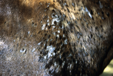
Hyperpigmentation (hypermelanosis), or melanoderma, is associated primarily with increased melanin in the epidermis and corneocytes. Histologically, there may also be dermal pigment, but when the majority of the pigment is in the dermis, a slate or steel blue coloration is present. Hyperpigmentation is frequently encountered as an acquired condition, usually associated with chronic inflammation and irritation.1,5 Hyperpigmentation may affect only the skin (melanoderma), only the hair (melanotrichia), or both of these. Melanoderma is often difficult to appreciate in dark-skinned horses and is most commonly seen with chronic hypersensitivity disorders. Melanotrichia is a common response when hair regrows following inflammatory insults (Fig. 12-1). The epidermis overlying numerous neoplasms and granulomas can become hyperpigmented.
Lentigo (plural, lentigines) is an idiopathic macular melanosis of horses.5 Annular, nonpalpable, dark black spots appear, usually with age, especially on the muzzle, on mucocutaneous junctions, and in the inguinal areas. The lesions are asymptomatic and of only cosmetic significance. Lentigo can be seen in association with melanocytic neoplasia in horses (see Chapter 16).
Hypopigmentation
Hypopigmentation (hypomelanosis) refers to a decrease of pigment in the skin of hair coat in areas that should normally be pigmented. Amelanosis (achromoderma, achromotrichia) indicates a total lack of melanin. Depigmentation means a loss of preexisting melanin. Leukoderma and leukotrichia are clinical terms used to indicate acquired depigmentation of skin and hair, respectively.1,3,5 Hereditary hypomelanoses have been divided into melanocytopenic (absence of melanocytes) and melanopenic (decreased melanin) forms.1 Pigment loss may occur from melanocyte destruction, dysfunction, or abnormal dispersion of melanosomes due to abnormal melanosome transfer or from inflammation. Lack of or decrease in melanosome production may also be a cause. The disorder may be congenital or acquired.
Genetic
Albinism
Albinism is a hereditary lack of pigment that is transmitted as an autosomal recessive trait.1,3,5 Albino individuals have a normal complement of melanocytes, but they lack tyrosinase for melanin synthesis and thus have a biochemical inability to produce melanin. Therefore, histopathologic studies reveal a normal epidermis with no pigment, but clear basal cells representing melanocytes are still seen. Skin, hair, and mucous membranes are amelanotic. Animals may also have unpigmented (pink) irides and photophobia.
Waardenburg-klein syndrome
Waardenburg-Klein syndrome has been described in American paints.1 In addition to blue eyes and amelanotic skin and hair, the affected animals are deaf and have blue or heterochromic irides.1,5 The defect is in the migration and differentiation of melanoblasts. Therefore, the affected skin has no melanocytes present. The syndrome is transmitted as an autosomal dominant trait with incomplete penetrance, so these animals should not be used for breeding.
Lethal white foal syndrome
Lethal white foal syndrome (overo lethal white foal syndrome, ileocolonic aganglionosis, myenteric aganglionosis, congenital intestinal aganglionosis) is an autosomal recessive disorder which is primarily a problem in paint horses, especially in overo breedings.*
In paints, there are three main coat patterns: overo, tobiano, and tovero. Genetic testing revealed that 95% of frame overos, 58% of toveros, and 100% of tobianos were heterozygous carriers.6a The defective gene was also found in the American miniature horses, half-Arabians, Thoroughbreds, and cropout Quarter horses.4a,6a Genetic testing is available through commercial laboratories.
Coat color dilution lethal
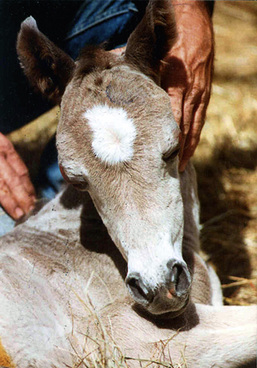
Coat color dilution lethal (lavender foal syndrome) is a rare hereditary condition of newborn Arabian foals of Egyptian breeding.5,12,12a The foals are born with a diluted or bleached-out hair coat color varying from a striking iridescent silver to pale lavender hue (“lavender foal syndrome”) (Fig. 12-2), to pale slate gray (“pewter”), to pale chestnut (“pink”).12 In addition, the foals have various neurologic abnormalities (unable to assume sternal recumbency, opisthotonus, paddling, extensor rigidity, seizures, blindness). The condition is fatal. Necropsy examination reveals no lesions.12
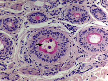
Histopathologic abnormalities in skin-biopsy specimens are characterized by dysplastic hair shafts and abnormal clumping of melanin in hair follicles and hair shafts (Fig. 12-3).5,12.


