Chapter 57Physitis
Physitis occurs in the horse in three principal forms: infectious physitis and type V and type VI Salter-Harris growth plate injuries.1 Type V growth plate injury can arise secondary to congenital but persistent angular limb deformities in foals, but these, infections, and type VI injuries are not considered in this chapter. Type V Salter-Harris injuries are considered a manifestation of acquired physitis, one of the group of disorders known as developmental orthopedic disease.2 The inclusion of acquired physitis under the umbrella of osteochondrosis may be more debatable. Jeffcott3 considered that the condition was better defined as physeal dysplasia, implying a disturbance of endochondral ossification rather than an inflammatory condition. Bramlage4 outlined two forms of physitis based on radiological interpretation of the site at which changes were seen. The two forms were those that were seen at the periphery, usually on the medial aspect, and those that were seen in the axial or central section of the growth plate.
Physitis is largely confined to the lighter-boned, faster-growing breeds of horse, particularly Thoroughbreds and Thoroughbred crossbreeds. Milder physitis is relatively common, and rarely does experienced stud management allow the condition to develop far enough for severe lameness and conformational changes to occur. Only in horses with more extreme or persistent physitis would radiographic examination be undertaken in the United Kingdom. Clinical recognition of enlargement of the growth plate or slight change in conformation usually results in a presumptive diagnosis of physitis and indicates an immediate change of the horse’s management as the first line of treatment.
Pathogenesis
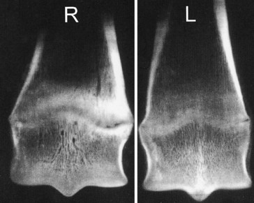
I consider the pathogenesis of physitis to be consistent with a type V Salter-Harris growth plate injury. This implies a compression lesion that arises medially as a consequence of the greater weight being borne on the medial aspect of the forelimb. Although the medial aspect of the forelimb is the most frequent site, physitis also occurs laterally in a foal with a carpal valgus deformity. Physitis arises at a time in the most active growing phases of young horses, when endochondral ossification is at its peak in the affected physis. It also occurs in the contralateral limb after a chronic lameness, at the fetlock in a younger foal, or at the carpus in older foals and yearlings. For example, I have encountered physitis in the contralateral forelimbs of foals with severe acquired flexural deformity of the distal interphalangeal joint (Figure 57-1). Although physitis may result in an upright conformation of the fetlock joint, probably from off-loading the limb to relieve pain, physitis does not cause other flexural deformity.

Full access? Get Clinical Tree

