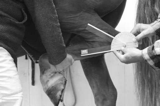
Many therapies are claimed to stimulate the regeneration process, often by a reduction of inflammation and swelling, an increase in blood circulation and alleviation of pain. Depending on the aim of the treatment and its specific target tissue, its efficacy may be measured with a variety of assessment tools, often with higher intra-rater reliability than inter-rater reliability. For example, pain perception can be assessed with palpation1, pain scales and questionnaires2, muscle tenderness (mechanical nociceptive threshold) with an algometer3,4, the degree of swelling and muscle bulk can be measured with diagnostic ultrasound, but also with simpler tools like measuring tape and calipers. The joint range of motion may be assessed with goniometer (Fig. 64.1).5 Lameness can be evaluated by ordinary clinical lameness examination, but also with different forms of motion analyzes such as force plates, pressure mats, and high-velocity filming.6 It is proposed that by decreasing tissue temperature, cryotherapy can reduce pain, tissue metabolism, muscle spasm, and minimize the inflammatory process, thereby stimulating tissue regeneration. Tissue temperature of 10–19°C may be needed to optimize these therapeutic effects. However, temperatures below 10°C may lead to tissue injury.7,8 After an injury, reduced hemorrhage and subsequent swelling is explained by a reduction in blood perfusion due to vasoconstriction. A significant decrease in soft tissue perfusion of the equine digit has been demonstrated scintigraphically in feet subjected to cryotherapy for 30 minutes.9 It is well recognized that cryotherapy can prevent swelling from occurring, but it remains debatable whether it may help reduce edema that is already present.7,8 A decrease in inflammation is also explained by decreased local metabolism and enzyme activity. It is suggested that tissue temperature needs to be around 10°C for a period of about 15 min in order to reduce skin metabolic rates11 and a joint temperature of 30°C to slow the cartilage degrading enzymes.12 An analgesic effect is due to a reduction in nerve conduction velocity, and a reduction in muscle spasm caused by the influence on muscle spindle fiber activity.13 It is shown that cold water immersion had the greatest effect on human tissue temperature and sensory nerve conduction.14 Further, it is hypothesized that cryotherapy is working through the gate theory with intense stimulation of cold sensitive receptors at spinal level. There are also indications that cryotherapy reduces muscle force and affects proprioception, through a reduction in firing of muscle spindles and Golgi tendon organs. As a negative effect, it is recognized that collagen becomes less pliable with cold, along with a subsequent temporary reduction in mobility.7 Cryotherapy should be started immediately after the injury and continued until the response to trauma is stabilized, usually 24–72 hours. The general recommendation is that cold should be applied for at most 20 minutes, every two hours. Based on review articles in humans, recommendations range from 20 minutes 2–4 times per day, up to 30–45 minutes every two hours.15 LaVelle & Synder suggest that 10 rather than 20 minute intervals achieve the same skin temperature, but with less ‘hunting response‘ (an axonal reflex causing a secondary vasodilatation that is believed to occur after 20–30 minutes of cold treatment).16 However, the question of a hunting response is still debated, and probably requires target areas that involve muscles. There are indications that cryotherapy does not create hunting responses in the treatment of equine distal limbs.9,17,18 Using shorter repeated cold applications allows the skin temperature to return to normal between treatments, thus maintaining the low muscle temperature without endangering the skin. In horses, the thermal response to cryotherapy has been studied in skin,17,19 tendons20,21 and hoof.9,18 Cold water immersion is generally recommended at a temperature of 14–16°C. Ice water immersion (0°C) may be a more effective way of cooling the distal limb than cold packs, cooling splints and crushed ice. In humans, application of a pack of frozen peas produced skin temperatures adequate to induce skin analgesia in 10 minutes, and reduced metabolic enzyme activity to clinically relevant levels in 20 minutes. Finally, the application of cold should be monitored carefully to avoid ice burn. A damp barrier (like a wet towel) can be used between a contact cold source and the skin. There are studies in horses that support the clinical efficacy of cryotherapy. Studies indicate positive effects of 5–9°C cold water treatment in horses with digital flexor tendon damage and suspensory ligament injury22, as well as a positive effect on laminitis.23 Another study showed a temperature decrease of 21°C after the application of cold, reaching a minimum tissue temperature of 10°C.21 Finally, ice water immersion of the distal limb, at 3°C for 48 hours, did not adversely affect the tissue.18 Most studies on cryotherapy in humans have used pain as an outcome measure. However, the efficacy of cold on relieving pain in clinical settings is uncertain. A Cochrane review reports that that ice alone seems to be more effective to reduce pain and swelling than control.15 Further, the application of ice immediately before a rehabilitation program significantly decreased pain and increased weight bearing.24 Ice therapy has showed benefit on pain relief, edema, joint motion and function, compared with control.10 It is proposed that by increasing tissue temperature, heat therapy can reduce pain, muscle spasm, joint stiffness and edema. The analgesic effect is explained by a direct influence on nerve conduction.7,8 Although superficial heat may only reach the superficial parts of tissues (approximately 1 cm from the skin surface), there is a possibility that it affects superficial nerves and thus have both a sedative and pain-relieving effect explained by the gate control theory. A reduction in muscle spasm is explained by the influence of heat on muscle spindle fiber activity, breaking the pain-muscle spasm-pain cycle. It is claimed that temperatures over 42°C slow the signaling rate of muscle spindles, but increases the signaling of Golgi tendon organs.7 Heat also reduces the viscosity of soft tissues and increases the elasticity of scar tissue, as well as reducing joint stiffness.24 Reduction of edema is explained by a vasodilatation effect, through spinal reflexes leading to a decreased smooth muscle tone of vessels. The increase in tissue temperature also creates an increase in metabolism. Enzymatic reactions are believed to increase with temperatures over 45°C, and a rise of 2–5°C is claimed to increase the action of phagocytic cells and cause an increase in blood flow with a reduction in swelling.7,8,12 Superficial heat can be applied with different types of heat packs covered in a moistened towel. The heat packs usually stay warm for approximately 30 minutes. Temperatures in superficial tissue can increase approximately 5°C after six minutes of superficial heat, and it takes approximately 15–30 minutes for deeper tissues to reach therapeutic levels. After approximately nine minutes of hot water hose therapy at a temperature of 38–41°C, the skin temperature over the equine metacarpal region was raised to 39.5–41°C and the subcutis to 39–40°C.20 Further, tissue temperatures return to baseline approximately 15 minutes after end of treatment. As previously mentioned, heating of connective tissues before stretching facilitates joint motion. To maintain tissue flexibility the temperature must be raised to 40–45°C for 5–10 minutes. Thereafter, the tissue must be put under stress.24 Studies in horses have shown a rise in tissue temperature after treatment with gel wraps, warm water hose and warm water bath.9,17,20 A 25 % increase in blood flow was reported after a 30 minute, 47°C water bath.9 A non-controlled study on 102 horses with various injuries reported a positive effect of dry whirlpool therapy, and that the temperature of the subcutis was raised 5°C.25 In humans, there are no conclusive data on superficial heat therapy for either swelling or pain.15 The mechanisms of actions for therapeutic ultrasound are divided into thermal and non-thermal effects.26 The thermal effects include an increase in blood circulation,27 and a direct influence on the nerve through a prolonged nerve transmission velocity.7 A non-thermal effect is explained by vibrations causing ‘micro-massage at the cell level‘, which is thought to increase the cell membrane potential, permeability, and transport mechanisms. The vibrations are also believed to create micro-currents of liquid and cavitation (the formation of tiny gas bubbles in the tissues as the result of ultrasound vibration), which may cause tissue destruction.7 The eventual effect of therapeutic ultrasound on bone healing is explained by an enhancement of the endochondral portion of fracture healing. Other proposed non-thermal effects are the acceleration of the inflammatory phase, stimulation of fibroblast proliferation and decreased pain.7,27,28 It is important that the electric apparatus and cords are functioning and protected from damage. Before using the ultrasound, one should make sure that it works properly by covering the transducer head with gel (or put it in a water bath) and increase the wattage to check if ‘bubbles‘ are created. It is also important to calibrate the instrument regularly, since studies have shown a variability of 30% in output intensity.28 In horses, treatment is usually performed at 1.5 W/cm2 for 10 minutes, once or twice daily for 10–14 days. The thermal effects of ultrasound are difficult to predict because of the varying rates of absorption by different tissues. A lower temperature rise was seen in the treatment of dogs with an intact hair coat, compared to dogs with clipped hair. However, there was a great temperature increase within the hair. Temperature increase of 0.6–3.5°C has been shown at 1–10 cm depth in canine muscles after treatment with 1–2 W/cm2 and 1–3 MHz.26 Lippiello and Smalley draw the conclusion that bone healing in equines was stimulated by ultrasound therapy.29 There are divergent results from studies on muscle and tendon regeneration in horses and dogs.30–34 In a clinical study without case controls, Lang treated 53 horses and 143 small animals. Based on owner evaluation, 63 % of the treatments were successful.35 One study failed to demonstrate any positive effects of ultrasound therapy on the healing of surgically severed Achilles tendons in dogs (1 MHz, pulse ratio1 : 4, 0.7–1.0 W/cm2, 10 minutes, three times a week, 13 treatments). In human systematic reviews authors conclude that there is limited evidence to support the use of active ultrasound therapy for treating people with musculoskeletal disorders.36,37 Contraindications are infectious arthritis, neoplastic diseases, and acute unstable fractures. ESWT should not be administered over the lung field, head, heart, major blood vessels or pregnant uterus. Adverse reactions such as pain, increased lameness, hematoma and inflammation have been reported in horses. Studies on focused ESWT show a disorganization of collagen network in the treated normal tendon38, as well as a potential to increase bone microcracking39. These studies recommend restricted exercise after treatment. However, other studies with ESWT on bone and bone-ligament interface showed no damage. The mechanism of action of ESWT is associated with the mechanical tension and pressure that the waves induce on tissue. Shockwaves release energy at the interfaces between tissues. It is proposed that ESWT increases blood circulation and stimulates the alignment of fibers in tendons and ligaments. Normal tendons treated with ESWT showed a transient stimulation of metabolism in tendinous structures, but six weeks after treatment the metabolism and level of glucosaminoglycans was lower than before treatment.38,39
Physical treatment of the equine athlete
Introduction
Thermotherapy
Cryotherapy
Background
Mechanisms of action
Therapeutic protocol
Outcome measures
Heat therapy
Background
Mechanisms of action
Therapeutic protocol
Outcome measures
Therapeutic ultrasound
Mechanisms of action
Therapeutic protocol
Treatment regimes
Outcome measures
Extracorporeal shockwave therapy (ESWT)
Indications and contraindications
Mechanisms of action
![]()
Stay updated, free articles. Join our Telegram channel

Full access? Get Clinical Tree


Physical treatment of the equine athlete

