23 Persistent primary teeth
ORAL EXAMINATION – UNDER GENERAL ANAESTHETIC
In summary, examination under general anaesthesia identified the following:
2. Persistent primary teeth
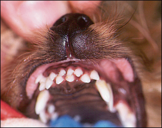
• The primary upper incisors were solidly (no mobility) in place and the permanent counterparts were erupted caudal to them (Fig. 23.1).
• The primary lower incisors had been shed and the erupted permanent lower incisors were occluding between the persistent primary and the permanent upper incisors (i.e. reverse scissor occlusion of permanent incisors).
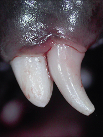
• All four primary canines were solidly in place. The diastema between the third upper incisor and upper canine was filled by the permanent upper canine erupting in front of the persistent primary canine bilaterally (Fig. 23.2). The permanent lower canines were trapped medial to the persistent primary canines, and were occluding with and traumatizing the palatal mucosa.
Stay updated, free articles. Join our Telegram channel

Full access? Get Clinical Tree


