Pediatric Fracture Management

CHAPTER 25 Pediatric Fracture Management


Fractures encountered in juvenile animals differ from those in adults in several ways. This chapter outlines a systematic method of pediatric fracture assessment, which can guide decision making regarding fracture management. Specifics regarding the treatment of physeal fractures and the use of external coaptation in pediatric patients are discussed. It is not intended to be a comprehensive review of internal fixation or external coaptation. The reader is referred to other textbooks for in-depth discussion of these topics.


The distinct characteristics of pediatric fractures result from the anatomic, biomechanical, and physiologic properties of pediatric bone. Anatomically, perhaps the most obvious difference between adult and pediatric bone is the presence of a growth plate, or physis. Physeal injuries are unique to immature patients. Biomechanically, pediatric bone is able to withstand greater plastic deformation before breaking than adult bone. This results in the classic incomplete “greenstick” fracture, not typically seen in adults. In addition, the presence of a strong periosteum minimizes displacement of fracture ends, even in cases of complete fractures. Physiologically, the robust periosteum and an already activated osteogenic environment result in rapid bone formation and accelerated healing of fractures. These differences in fracture configuration, healing potential, and physical characteristics of bone in pediatric patients dictate a unique approach to fracture management.


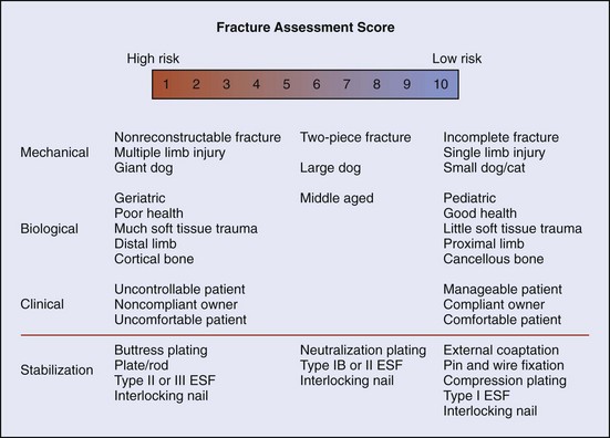
Fracture Assessment Score


Fracture management has been described as a race between biologic healing of the fracture and mechanical failure of the stabilization used. The more mechanically or biologically favorable the fracture environment, the less robust or long lived need be the stabilization. The fracture assessment score is an objective method of considering all pertinent fracture characteristics to aid in fracture management decision making. It evaluates mechanical factors, biologic factors, and clinical (patient/client) factors and rates them on a scale of 1 to 10 (Box 25-1). Higher scores are given for factors that predict faster healing, minimal loads on implants, and decreased risk for implant failure. In general, pediatric patients tend to have higher scores for mechanical factors and biologic factors, predisposing them to more rapid healing. However, clinical factors may score lower than in adult patients. An overview of the scoring system is presented in Figure 25-1.


When using the fracture assessment score, it is not critical to come up with an absolute number, as this will probably vary between observers. The important point is that all three factors are considered when making decisions regarding fracture management. Because mechanical factors are the most obvious, and the most easily visualized on the radiograph, the most common error in fracture assessment is to ignore the biologic and clinical factors. This can lead to failure of a fixation that may have appeared appropriate when reviewing radiographs alone.


Mechanical factors that affect bone healing include fracture configuration, patient size, and presence of single limb versus multiple limb injury. This predicts the ability of the fractured limb to share load bearing with the fixation device, as well as the magnitude of load that will be exerted on the limb. Pediatric patients tend to present with a higher proportion of incomplete fractures and simple fractures with contact of fragment ends. These configurations have a higher load-sharing ability than comminuted, nonreconstructable fractures. Thus they have a correspondingly higher assessment score. Larger dogs or dogs with multiple limb injuries can be expected to exert larger forces on the fracture at earlier time points. They will have a lower assessment score than small dogs with single-limb injuries. In general, the mechanical assessment dictates the initial strength of implant needed to stabilize a fracture.


Biologic factors that affect the rate of fracture healing include age, health, soft tissue injury, and region of bone affected. Because they are “physiologically primed” to produce bone, pediatric patients in good health exhibit significantly accelerated bone healing (higher assessment score) compared with adults or geriatric patients. Fractures resulting from high-velocity injury or open fractures often result in tremendous soft tissue trauma. Soft tissue injury in the fracture region will compromise blood supply to the bone, decreasing healing potential and indicating a lower assessment score. Bones of the distal extremities (radius and tibia) are surrounded by a smaller volume of muscle and soft tissue. Consequently they have diminished extraosseous blood supply and exhibit an increased rate of delayed fracture union, even in pediatric patients. Fractures involving cancellous bone tend to heal more rapidly because of the presence of a large number of osteoblasts and osteoinductive factors compared with cortical bone.


The increased healing potential of growing animals is a great asset in pediatric fracture management. Sometimes this may tempt the surgeon to compromise some mechanical principles of fracture fixation for reasons of expense, available equipment, or time. However, the biologic environment alone cannot compensate for inadequate mechanical fracture stabilization, and the contribution of both factors must be acknowledged when choosing fixation technique. The biologic assessment predicts the required duration or functional lifespan of the stabilization. Because larger implants have a longer fatigue life, a lower biologic assessment score may indicate use of larger implants. The opposite is true for a high biologic assessment score.


Clinical factors that affect bone healing include postoperative patient cooperation, patient comfort, and owner compliance. Uncontrollable, active patients tend to be poor candidates for external fixators or external coaptation and will be better served by internal fixation. Owners who are unable or unwilling to perform the postoperative management necessary for casts or external fixators are better off having their pet’s fracture managed with internal fixation. Early return to function is important with fracture management. For some dogs, the presence of an external fixator or coaptation will inhibit normal limb use. These dogs may be predisposed to complications such as loss of range of motion or muscle atrophy and may be better managed by internal fixation. Clinical assessment may alter the indicated strength or duration of fixation (as predicted by mechanical and biologic assessment), or it may dictate alternate management that is better tolerated by the patient or client.


The final fracture assessment score can be used to guide fracture management. It is most applicable for diaphyseal fractures, for which many options for stabilization exist. In other applications, such as articular fractures or physeal fractures, the mechanical environment may largely dictate the repair, and there may be little choice in management options.


Physeal Fractures


Fractures of the physeal plate are not uncommon in young animals. The zone of hypertrophy in the physis is a focal weak point in the long bone. Ligaments and connective tissue are significantly stronger than the physis in pediatric patients. Thus trauma that would typically result in a sprain or luxation in adults can cause fracture or displacement involving the physis. Physeal fractures are categorized according to the method of Salter and Harris into types I through V, as described in Chapter 42 (see Figure 42-1), primarily in an attempt to prognosticate outcome.


The Salter-Harris system is useful for describing most physeal fractures; types I through IV can usually be differentiated radiographically. However, predicting outcomes for physeal fractures is more complex, and the Salter-Harris system is limited in this regard. Whereas the articular nature of types III and IV fractures indicates a worse prognosis than type I or II (as a result of the development of degenerative joint disease), the difference in prognosis between types I and II or types III and IV is less clear cut. Type V fractures probably have the worst prognosis because injury to the germinal cell layer of the physis can result in arrested growth and development of angular deformities or length deformities. However, the crushing injury of type V fractures cannot be visualized radiographically, making them very difficult or impossible to diagnose at the time of injury. Moreover, type V fractures often are not a distinct entity; many type I through IV fractures have some degree of type V damage to the growth plate. Any trauma significant enough to cause a fracture (physeal or otherwise) may also cause type V physeal damage to the fractured bone or to a separate bone.


Because of the occult nature of type V physeal fractures, all puppies and kittens with fractures should be reevaluated at least every 2 weeks to assess for the development of angular limb deformities, regardless of the method of treatment. Age at time of injury is a significant factor in prognosis for physeal fractures. Younger animals are at increased risk for growth deformities, and those less than 6 to 7 months old should be monitored more closely. If deformities are diagnosed early, residual growth potential in the limb can be used to aid in treatment. Damage to the physeal plate is of lesser consequence in animals more than 8 to 10 months of age because less growth potential remains.


Sep 11, 2016 | Posted by in SMALL ANIMAL | Comments Off on Pediatric Fracture Management

Full access? Get Clinical Tree

Get Clinical Tree app for offline access