Pathophysiology and Clinical Diagnosis of Cortical and Subchondral Bone Injury

Chapter 100Pathophysiology and Clinical Diagnosis of Cortical and Subchondral Bone Injury


Pathophysiology


Bone is an amazing living tissue that is able to conform and adapt to its environment. When bone is exposed to a load, it deforms. This deformation is elastic and within certain limits; bone returns to its original state once the load is removed. If the degree of deformation is beyond bone’s upper limit, complete failure occurs. Under other conditions, bone may be subject to loads that are persistently different. Alterations in bone strain patterns develop, and the bone responds accordingly. Bone will then counteract the variation in load by changing its inertial properties via the processes of modeling and remodeling. In time, bone will modify its shape and structure, effectively adapting to the load under which it is placed (Wolff’s law).


The term modeling refers to the geometric sculpting of bone by formation or resorption.1 With modeling, osteoblasts and osteoclasts work independently, resulting in increased or decreased bone density. Resorption occurs when strain patterns are below the minimum threshold, and bone formation occurs when bone strain is above the maximum strain threshold.1 In cortical bone, modeling adaption results in periosteal thickening (bone formation) or thinning (bone resorption). In trabecular bone, subchondral sclerosis or osteoporosis ensues. Eventually, modeling alters the size, shape, and density of bone. Remodeling refers to the coordinated action of osteoclasts and osteoblasts resulting in the removal of biomechanically inferior bone and replacement with new bone.1 Osteoclasts move through bone at a much quicker rate than osteoblasts; therefore bone resorption (within weeks) is followed by bone formation (months).


Bone modeling and remodeling are active ongoing processes and can occur in the same bone at the same time. These processes are driven by site-specific bone strain patterns, although the exact mechanism by which strain induces bone change is unknown. Throughout life, bone continually adapts to its physiological loads by modeling and/or remodeling until its mechanical stimulus is normalized. These processes ensure the mechanical integrity of the skeleton. For example, when a horse undergoes athletic training, its bones are exposed to increasing and varying loads at specific locations. In turn, bone responds by adding more bone to regions of higher load. An adaptive site-specific response to race training in Thoroughbred (TB) horses is the dramatic thickening that occurs in the dorsomedial aspect of the third metacarpal bone (McIII).


In ideal situations, a balance exists between load and the rate of bone adaptation. Repetitive cyclic loading produces an accumulation of focal microdamage at sites that are maximally stressed. Microdamage incites a bone response, followed by successful repair and remodeling. If there is imbalance between removal and replacement, the very process of bone repair may contribute to a bone’s failure. Because osteoclasts act more quickly than osteoblasts, transient weakness occurs after damaged bone has been removed and before completion of bone replacement. Prolonged or rapid increases in load, such as those during high-speed workouts and racing, result in an accumulation of microdamage. The resorptive phase of remodeling may then exceed replacement capacity and transiently weaken the bone.2,3 This focal weakness can function as a stress riser and allow initiation of a complete fracture or fragmentation under otherwise normal physiological loading.4


Failure of the bone to adapt in a timely fashion combined with continual and compounded microdamage results in stress-related bone injury.5 Horses undergoing race training and racing are particularly susceptible to injury because their bones are constantly subjected to large repetitive loads. Risk for injury is high until a satisfactory adaptive bone response is completed.3 Evaluation of postmortem specimens indicates that most musculoskeletal injuries are overuse injuries. Evidence of stress remodeling has been observed in equine long bones (the McIII, humerus, scapula, or tibia), the third carpal bone (C3), the pelvis, and vertebrae.6-14 Site-specific pathology includes periosteal callus, sclerosis of endosteal or subchondral bone, and focal osteoporosis.


Stress-related cortical bone injury is commonly referred to as a stress or fatigue fracture. Stress fractures occur in horses undergoing intense exercise (repetitive high-strain loading) and are not correlated with a specific traumatic event.3,8,15-17 They are associated with activities that produce repetitive loading of the involved bone. Repeated cyclic loading of cortical bone results in loss of stiffness, reduction in strength, and development of microcracks. With continued loading, microcracks propagate and coalesce into macrocracks (i.e., stress fractures). Stress fractures are regularly identified, clinically and at postmortem examination, in consistent locations presumably at sites of maximal load. They are very common in racehorses and are a well-recognized cause of lameness. If the stress remodeling continues in an unbalanced fashion, trauma to an already fatigued bone can lead to catastrophic fracture. Periosteal callus, indicative of preexisting reparative response, at equine cortical fracture interfaces6,13 is common and provides evidence of a continuum of stress-related bone injury.


Like cortical bone, subchondral bone is susceptible to stress-related bone injury. Although features of bone modeling and remodeling are similarly induced, depending on the mechanical impact periosteal callus is not seen. Normally, the compliant subchondral plate acts as a shock absorber between articular cartilage and subchondral bone, thereby dissipating the impact to the articular surface. Repeated loading results in subchondral bone mineralization and subsequent stiffening to combat the increased bone strain. The progressive increased bone density of the trabecular bone adjacent to the subchondral plate of the radial fossa of the C3 and the palmar condyles of the McIII or plantar condyles of the third metatarsal bone (MtIII) are examples of a subchondral response in race-trained horses.12,18,19 As with cortical bone, repetitive loading of the subchondral bone may result in a repair process dominated by the resorptive phase of remodeling. Associated focal subchondral osteoporosis and microcracks may result. Small cracks may develop into larger fractures, resulting in fragmentation and overt fracture along articular margins.20 Preexisting subchondral sclerosis is commonly seen in racehorses with distal McIII condylar fractures21 and is a proposed precursor to C3 slab fractures.12,22 Sclerotic subchondral bone may result in articular cartilage damage. Progressive cartilaginous erosion and ulceration has been identified at areas of increased subchondral thickness.9,23,24 Collapse of subchondral bone resulted in flattening and indentation of cartilage, osteochondral fragmentation, and ultimately osteoarthritis (OA).7,9,20


Clinical Examination


A pathological continuum of cortical and subchondral stress-related bone injury causes lameness, subchondral sclerosis, subchondral lucency, incomplete (stress or fatigue) and complete fracture, and, in horses with subchondral damage, the eventual development of OA. Clearly, early and accurate diagnosis of stress-related bone injury in racehorses is important for the well-being of the horse and the safety of the industry. However, clinical recognition of these injuries can be challenging, and, although stress fractures are a well-recognized cause of lameness in racehorses,11,15,16,25-34 most of these injuries occur in the absence of a specific traumatic event.3,8,15-17,30 Characteristic history includes acute onset of lameness after racing or training that responds to rest.25-27,30,35 Over time, lameness may worsen or linger, and some horses may have a history of weeks to months of poor performance, intermittent unilateral lameness or lameness in numerous limbs, or reports from exercise riders or drivers that horses are not “right.”27,36,37 Lameness scores at the time of diagnosis are quite variable, with some horses exhibiting no lameness and others being severely lame. Physical findings such as swelling or sensitivity to palpation are often subtle or absent. This is especially true in horses with upper limb long bone stress fractures, in which palpation may be at best difficult.


Clinical signs of cortical bone injury include periosteal thickening and local sensitivity. This is easily recognized in horses with the bucked-shin complex38-40 (see Chapter 102). However, in the majority of long bones, overlying soft tissue and muscle prohibit clear identification. Localized signs of cortical pain in the tibia or humerus are uncommon and frequently absent.16,25,28,31 When noted, pain during palpation and percussion of the medial diaphysis of the tibia occurs in as few as 30% of affected horses.25 Careful palpation may reveal focal pain in horses with avulsion fracture of the proximal palmar or plantar aspect of the McIII or the MtIII, respectively.41 Pain on palpation or profound muscle spasm of the affected side and ventral displacement of a tuber sacrale are consistent but subtle physical abnormalities in horses with stress fractures of the wing of the ilium.15,30 Flexion tests, time-honored clinical tools to help exacerbate and localize lameness, often have inconclusive results, and findings may even be absent in horses with cortical bone injury.24,42


Clinical signs of subchondral bone injury are also variable and often subtle. Injuries are frequently bilateral, and overt unilateral limb lameness is unusual. Affected horses may simply be performing at a lower level than expected. Overlying cartilage is minimally affected early in the disease process, and joint effusion is absent. Early subchondral bone injury of the distal aspects of the McIII and the MtIII rarely causes fetlock effusion,36,43-45 and carpal effusion is often less than expected based on severity of lameness in Standardbreds (STBs) with subchondral lucency of the C3.46,47 Diagnostic analgesia is essential for accurate diagnosis, but perineural techniques are consistently more effective than intraarticular analgesia, presumably because pain is associated with subchondral bone and not synovitis, capsulitis, and overt cartilage damage (see Chapter 42). Veterinarians and trainers are often incredulous when a diagnosis of subchondral bone injury is made by use of diagnostic analgesia and scintigraphic examination when clinical signs are lacking. As with cortical bone injury, flexion test results are inconsistent and often negative in horses with subchondral bone injury.36,46 Subchondral increased radiopacity may be radiologically apparent, but it is often difficult to determine whether this is successful adaptive change or a maladaptive or nonadaptive response. In those horses with severe or chronic subchondral bone injury, clinical signs are often more obvious. Synovitis, lameness, and OA may be noted.


Diagnostic Analgesia


Although diagnostic analgesia is one of the most valuable tools used to localize the authentic source of pain, there are several considerations in horses with stress fractures and subchondral bone injury. At the time of evaluation, horses must be lame enough to visually assess response to diagnostic analgesia. Horses with stress fractures are notorious for being lame immediately after intense exercise but subtly lame or sound at the time of lameness examination. Neither perineural nor intraarticular analgesia can be used to diagnose upper limb stress fractures; however, such fractures should be suspected when lameness cannot be improved by routine distal limb diagnostic analgesia.* In addition, response to diagnostic analgesia is often variable. Lameness associated with cortical or subchondral injury of the proximal palmar aspect of the McIII may improve with high palmar analgesia or intraarticular analgesia of the middle carpal joint.27,41,42,50 Pain associated with nonadaptive subchondral remodeling of the distal aspect of the McIII or the MtIII may be alleviated by using a low palmar or plantar or the lateral palmar metacarpal or plantar metatarsal block.51 Often these horses will not improve with intraarticular analgesia and are similarly unresponsive to intraarticular medications.36,43,44 Diagnostic analgesia is a good starting point but must not be used alone.


Diagnostic Imaging


Radiography and Radiology


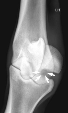
Radiography remains hugely important in the diagnosis of bone injury in racehorses; however, it is insensitive during early phases of stress-related bone injury and often inadequate for subtle subchondral changes. Radiology provides structural information, much of which must be several weeks old before it can be seen. Lag time for enough structural change to occur to be seen radiologically is an obviously serious limitation of radiography, especially when one is attempting to localize and identify early or subtle bone injury. It takes 2 to 3 weeks or more before radiological changes of periostitis are apparent (see Chapter 15).52-55 Even frank fracture lines or compression injury of subchondral bone can take days to weeks to be recognized. Supplementary radiographic images of site-specific regions may enhance detection of injury. For example, the dorsal 30° proximal 45° lateral-palmar (or plantar) distal medial oblique and dorsal 30° proximal medial-palmar or plantar distal lateral oblique (“down-angled”) images (Figure 100-1) are useful for evaluation of subchondral injury of the condyles of the McIII or the MtIII.51,55 A dorsal 60° proximal 45° lateral-palmar distal medial oblique image or dorsal 60° proximal 45° medial-palmar distal lateral oblique image of the distal phalanx is necessary for identification of palmar process fractures.56-58 Even with additional images, subtle subchondral radiolucency and increased radiopacity are often difficult to recognize because density differences must be at least 30% to 50% and lesions 1 to 1.5 cm in diameter before recognition is possible.52 Another obvious limitation of radiography is the inability to distinguish whether the radiologically apparent lesion is an active process or simply the result of an adaptive bone response.


Nuclear Scintigraphy


Increased awareness of location and prevalence of stress-related bone injuries and scintigraphic examination of affected racehorses enhance early detection of bone pathology before catastrophic fracture. Nuclear scintigraphy is an extremely sensitive method of detecting exercise-induced bone remodeling (see Chapter 19). Normal bone response in the dorsal aspect of the McIII, the proximal sesamoid bones (PSBs), and the metacarpal condyles is easily identified and has typical patterns of increased radiopharmaceutical uptake (IRU) in horses undergoing high-speed exercise.59,60 Scintigraphy is useful to detect abnormal alterations in local bone metabolism such as increased activity from cyclic loading and provides a foundation for the diagnosis of stress fractures, stress remodeling, and subchondral trauma.* A focal, moderate-to-intense area of IRU is the scintigraphic hallmark of stress fracture.


Several circumstances exist in which scintigraphy is invaluable for the diagnosis of cortical or subchondral bone injury. Racehorses with stress fractures often have a history of acute severe lameness that abates with rest. These horses have few localizing clinical signs and are diagnostic challenges. Scintigraphy can be used as a screening tool and obviates the need for numerous radiographic images. In addition, a negative bone scan is invaluable because it rules out active bone disease and virtually eliminates the possibility of a stress fracture.52,54 Another scenario is when a racehorse fails to respond to diagnostic analgesia. This is especially true in horses with upper limb stress fractures. Scintigraphy is also helpful for horses with bone injury in numerous areas or multiple limbs. Racehorses with stress remodeling of the distal aspects of the McIII or the MtIII are often in this category.


Scintigraphy is more sensitive than radiography for the identification of stress-related bone injury, and a positive bone scan may precede conclusive radiological change by at least 2 to 3 weeks.52-54 In reports of scintigraphic examinations of racehorses, nearly half of areas of IRU were not associated with radiologically detectable abnormalities.61,63,64 A positive bone scan may also enhance the recognition of subtle radiological changes41 and assist in determination of a structural abnormality as an active process or a reflection of past change. For example, radiographs of a 3-year-old TB racehorse reveal extensive thickening of the dorsal cortex of the McIII, but the bone scan is negative. Gradual resolution of IRU of the dorsal cortex of the McIII can be interpreted as evidence of satisfactory healing of a previously identified dorsal cortical fracture. On the other hand, a persistent fracture line without concurrent moderate-to-intense IRU may indicate the development of a nonunion, because the amount of radiopharmaceutical uptake reflects the rate of bony repair.29 Carpal pain is not likely in a STB with radiological evidence of extensive sclerosis of the C3 but with only mild or no IRU seen scintigraphically.


Ultrasonography


Ilial wing fractures can be detected ultrasonographically.15,17,30,66,67 Hematoma formation, irregular bone contour (indicative of callus formation), and breaks in the normal hyperechogenic contour of bone may be seen if the fracture involves the dorsal surface of the bone (see Figure 49-3, page 566). A displaced fracture is seen as hyperechogenic bony structure distracted from adjacent bone (“stair-step” sign). Ultrasonographic abnormalities are usually most severe at the caudal margins of the fracture.66,67 These findings are supported by postmortem studies in horses with ilial wing fractures.11,13 Serial ultrasonographic examinations can be used to determine if the fracture has healed but should not be the single means of diagnosis. Because of variability in conformation of the ilium and other factors, ultrasonographic findings should be combined with a thorough clinical examination and, if necessary, scintigraphic and occasionally radiographic examinations.15,66


Specific Locations of Cortical and Subchondral Bone Injury


Knowledge of the prevalence of stress-related bone injuries provides insight into the likelihood of an area causing clinically important problems, especially when diagnostic analgesia fails to localize the lameness, or radiological and ultrasonographic examination findings are unremarkable. The type of training and racing largely determines the location of injury and whether the injury involves cortical or subchondral bone (Tables 100-1 and 100-2). In lame STBs, IRU is most commonly associated with exercise-induced subchondral bone remodeling, and prevalent sites include the PSBs followed by the C3 and the tarsus.63 The distal palmar or plantar aspect of the McIII or the MtIII is also a frequent location of IRU and, when combined with IRU of the PSB, is the most common location of stress remodeling.55,63 In TBs, IRU of the distal palmar or plantar aspects of the McIII or the MtIII is the most common abnormal scintigraphic finding.62 In the forelimbs, sites of prevalence are the distal palmar aspect of the McIII, followed by the carpus and the dorsal cortex of the McIII.62 In the hindlimbs, the distal plantar aspect of the MtIII is the most common abnormal area, followed by the tarsus and tibia.62


TABLE 100-1 Prevalences of Sites of Increased Radiopharmaceutical Uptake in the Forelimbs of Racehorses


Only gold members can continue reading. Log In or Register to continue

Stay updated, free articles. Join our Telegram channel

Jun 4, 2016 | Posted by in EQUINE MEDICINE | Comments Off on Pathophysiology and Clinical Diagnosis of Cortical and Subchondral Bone Injury

Full access? Get Clinical Tree

Get Clinical Tree app for offline access
SITE STANDARDBRED (%) THOROUGHBRED (%)
Phalanges 14 10
Proximal phalanx 7
Middle phalanx 0
Distal phalanx 7
Proximal sesamoid bone 32 6
Third metacarpal bone    
Distal palmar 21 50
Dorsal 0 15
Proximal palmar 3 4.5
Carpus (C3) 43