, Patricia A. Noguera1 and Trygve T. Poppe2
(1)
Marine Scotland Science, Aberdeen, UK
(2)
Norwegian School of Veterinary Science, Oslo, Norway
Abstract
A methodical approach is a prerequisite for an accurate diagnosis and requires a description of the tissue changes that occur following infectious and non-infectious conditions. Cells have a limited repertoire of morphological response to injury which is linked to biochemical mechanisms that determine the outcome of cell damage, thus accounting for the appearance of cells within lesions inducing general pathological changes, rather than those that are pathognomonic. This chapter covers the different types of cell and tissue responses to acute or chronic injury.
Keywords
Fish diseaseInflammationProliferationCirculatory disturbancesNecrosisPigmentsNeoplasiaPathology is the study and diagnosis of disease, and the recognition and interpretation of the physiological and pathological processes. This requires a thorough understanding of normal tissue structure and microanatomy. Normal structure varies widely among species, age and physiological and developmental stages, and even within a population variations may occur, therefore understanding these changes and how they relate to the status of the species under investigation is essential. Furthermore, many diseases look similar at the gross level and to illustrate this aspect, images from skin, kidney and liver showing a range of lesions of different causes are presented in Figs. 4.1, 4.2 and 4.3.
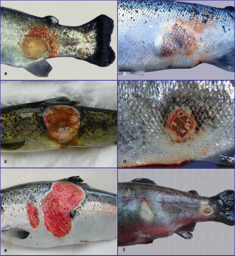
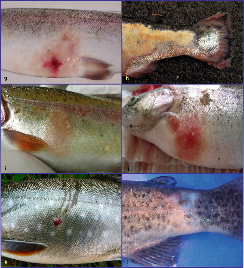
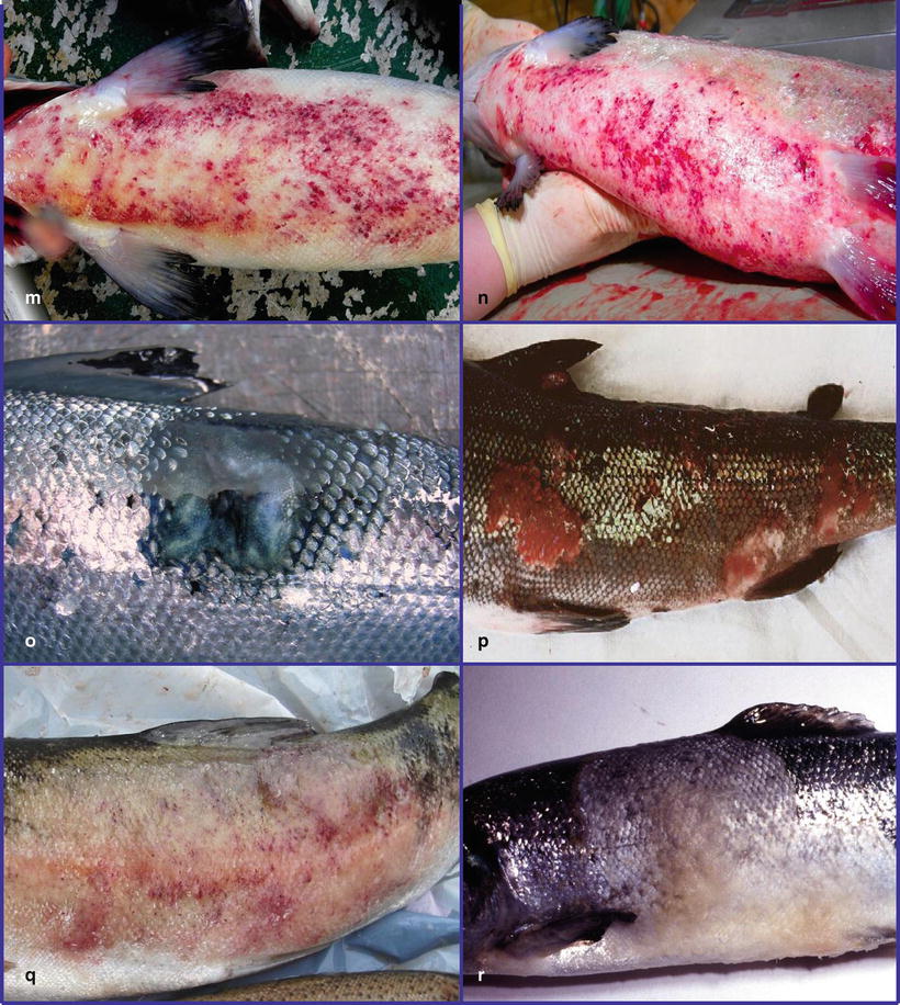
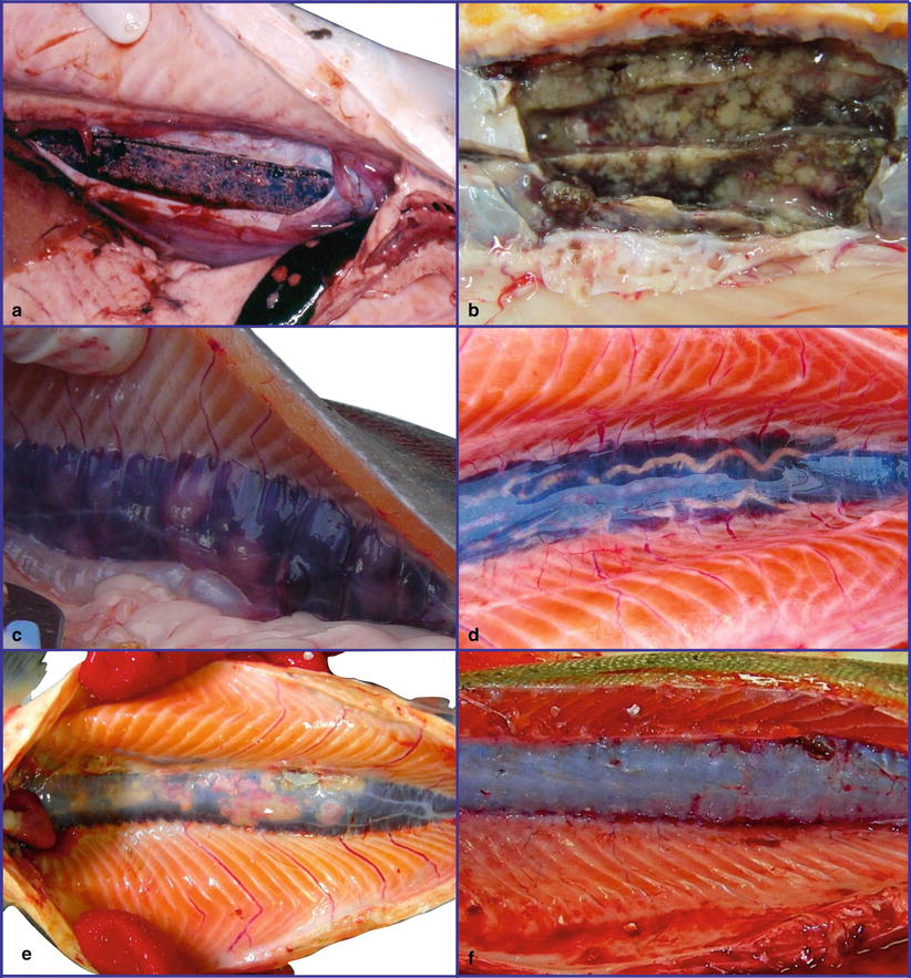
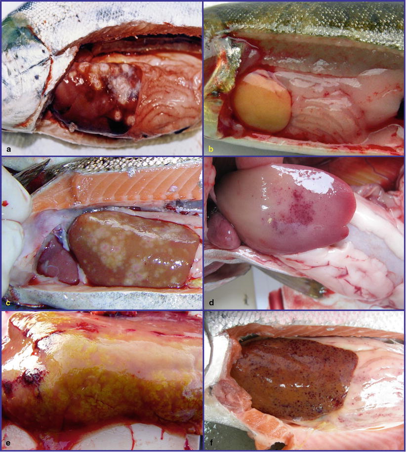
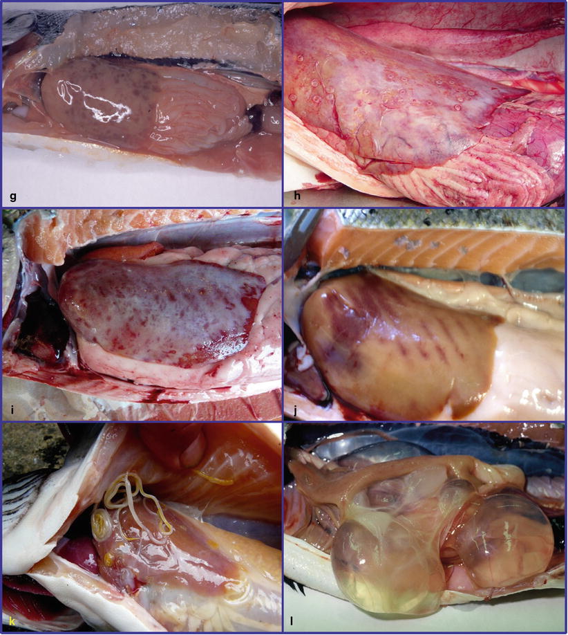


Fig. 4.1
(a) Skin ulcer caused by Aeromonas sp. in farmed rainbow trout. (b) Boil lesion caused by Aeromonas salmonicida subsp. salmonicida in farmed Atlantic salmon. (c) Deep skin ulcer caused by Pseudomonas fluorescens in Atlantic salmon smolt. (d) Healing Pseudomonas ulcer in Atlantic salmon. (e) Winter ulcer associated with Moritella viscosa in Atlantic salmon. (f) Skin ulcers caused by Flavobacterium psychrophilum in rainbow trout. (g) Classical Vibrio infection in Atlantic salmon. (h) Tenacibaculum maritimum in Atlantic salmon. (i) Early red mark syndrome in rainbow trout. (j) Advanced red mark syndrome in farmed rainbow trout. (k) Lesion associated with cormorant strike in Arctic char. (l) Peduncle disease caused by Flavobacterium psychrophilum in rainbow trout. (m) Ventral skin haemorrhage in Atlantic salmon with CMS. (n) Systemic Moritella viscosa infection in Atlantic salmon. (o) Healed skin ulcer in Atlantic salmon. (p) Papillomatosis in wild Atlantic salmon. (q) Puffy skin with haemorrhage in rainbow trout. (r) Saprolegnia infection in Atlantic salmon

Fig. 4.2
(a) BKD in Atlantic salmon. Kidney capsule has been opened. (b) Mycobacterium infection in Atlantic salmon. Kidney capsule has been opened (c) PKD in rainbow trout. (d) Phyllodistomum umblae in wild Arctic char. (e) Multiple necrosis caused by Spironucleus salmonicida in Atlantic salmon. (f) Non-specified kidney neoplasia in Atlantic salmon


Fig. 4.3
(a) Infection with Piscirickettsia salmonis in Atlantic salmon. (b) Pale, yellowish liver in Atlantic salmon smolt with Infectious pancreatic necrosis. (c) Multiple necrosis caused by Spironucleus salmonicida in salmon. (d) Enteric red mouth in rainbow trout. (e) Myxidium truttae plasmodia in bile ducts of wild Atlantic salmon. (f) Petechiae in septicaemic Atlantic salmon. (g) Haemorrhage caused by Listonella anguillarum in Atlantic salmon. (h) Anisakis simplex larvae in wild Atlantic salmon. (i) Fibrinous coat on liver in salmon with cardiomyopathy syndrome. (j) Post mortem artifact. (k) Philonema salvelini in wild brook trout. (l) Polycystic liver in Atlantic salmon
Cell types in fish are, in principle, the same as those found in mammals and similarly, many direct and indirect pathological stimuli induce general pathological changes rather than being pathognomonic. Cells have a limited repertoire of morphological response to injury and are linked to biochemical mechanisms that determine the outcome of cell injury, accounting for the appearance of cells within lesions.
A methodical approach is a prerequisite for an accurate diagnosis and this requires a description of the tissue changes that occur in relation to infectious and non-infectious agents, response to acute or chronic injuries, nutritional imbalance and other causes of disease or abnormality, followed by histopathology. The following areas will be covered in this chapter: inflammation, proliferation, circulatory disturbances, cell injury and necrosis, pigments and mineralization and neoplasia. Finally, a brief description of artefacts is provided to help distinguish these from tangible pathological changes.
Common prefixes and suffixes used in compounded words are provided in Table 4.1, and a glossary appropriate to veterinary terminology is provided in Chap. 13. Reference should also be made to Chap. 2 which discusses normal tissues.
Table 4.1
Examples of prefixes and suffixes and their use in compounded words
Prefix | Meaning | Example |
|---|---|---|
Adeno- | Glandular | Adenoma |
An- | No, not | Anaemia |
Angio- | Blood or lymph vessels | Angiopathy |
Anti- | Counteracting | Antibody |
Apo- | Separated from | Apoptosis |
Auto- | Self | Autoimmunity |
Cardio- | Heart | Cardiomyopathy |
Chol- | Bile | Cholangitis |
Con- | Together | Confluent |
Cyto- | Cell | Cytopathic |
De- | Remove or loss | Degeneration |
Derma- | Skin | Dermatomycosis |
Dys | Abnormal | Dysplasia |
Ect- | Outer or external | Ectoparasite |
Endo- | Within or inner | Endoparasite |
Enter- | The intestine | Enteritis |
Epi- | Above, upon | Epidermis |
Fibro- | Fibres or fibrous tissue | Fibroplasia |
Gastro- | Stomach | Gastrointestinal |
Haemo- | Blood | Haemolysis |
Hepato- | Liver | Hepatomegaly |
Hetero- | Difference | Heteropagus |
Histo- | Tissue | Histology |
Homo- | Similar, like | Homogenous |
Hyper- | Indicating an excess | Hyperpigmentation |
Hypo- | Indicating a deficiency | Hypoplastic |
Idio- | Self | Idiopathic |
Inter- | Between | Interstitial |
Intra- | Within | Intracellular |
Karyo- | Cell nucleus | Karyomegaly |
Leuco- | Lack of colour, white | Leucopenia |
Lipo- | Fatty | Lipoidosis |
Macro- | Large | Macrophage |
Mal- | Disorder or abnormality | Malignant |
Melan- | Black colour | Melanin |
Micro- | Small | Microcytic |
Morpho- | Structure | Morphological |
Multi- | Many | Multicellular |
Myco- | Fungus | Mycosis |
Myo- | Muscle | Myocardium |
Necro- | Death or dissolution | Necrosis |
Nephro- | Kidney | Nephrocalcinosis |
Osteo- | Bony | Osteoclast |
Patho- | Disease | Pathogen |
Peri- | Around or enclosing | Periorbital |
Phago- | Eat; devour | Phagocyte |
Post- | After | Posterior |
Poly
Stay updated, free articles. Join our Telegram channel
Full access? Get Clinical Tree
 Get Clinical Tree app for offline access

|