Pathogenesis of Osteochondrosis

Chapter 54Pathogenesis of Osteochondrosis


Osteochondrosis: Definitions and Terminology


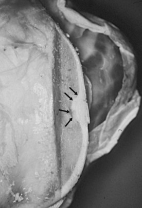
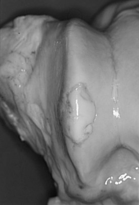
Equine osteochondrosis (OC) is characterized by focal failures of endochondral ossification that typically occur in well-defined predilection sites. Lesions that result from external trauma or infection are not generally regarded as OC.1,2 The thickened, retained growth cartilage that characterizes typical lesions (Figure 54-1) in the articular-epiphyseal cartilage complex (AECC) may be complicated by development of fissures that extend from the deepest layers of the lesion to the articular surface. Cartilaginous or osteochondral fragments may then detach from the parent bone, forming intraarticular fragments.3 Once lesions extend to the articular surface, thereby causing inflammation of the joint, the condition may be referred to as osteochondritis.4,5 The term osteochondritis dissecans (OCD) is usually reserved for lesions in which a dissecting flap of tissue is present (Figure 54-2).3 Although it has been proposed that the term dyschondroplasia should be used in place of OC,3,6 the term OC remains in widespread use and is used in this chapter to refer to the primary lesion. Dyschondroplasia is used only when referring to work by authors who prefer this term. A condition similar to equine OC occurs in a number of other animal species, as well as in people.1


Endochondral Ossification


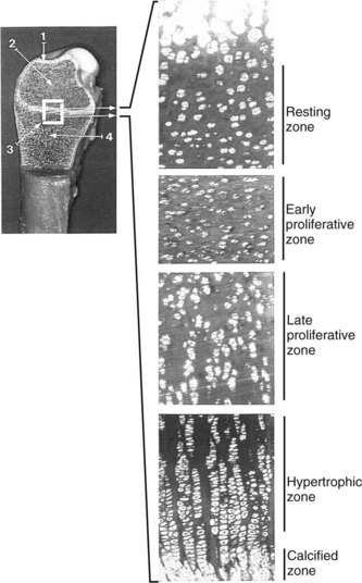
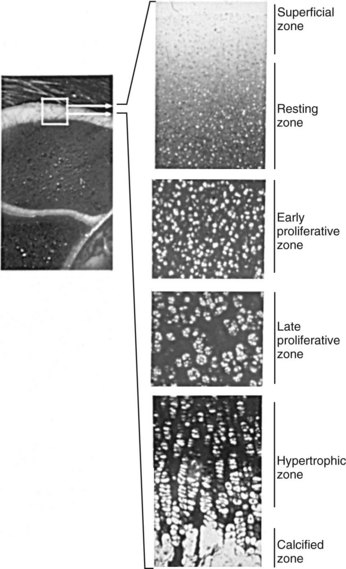
Endochondral ossification is the process by which growing cartilage is systematically replaced by bone to form the growing skeleton.7 This process occurs at three main sites: the physis, the epiphysis, and the cuboidal bones of the carpus and tarsus. Chondrocytes in the physis can be divided into a series of layers or zones (Figure 54-3). The zone farthest from the metaphysis is the resting or reserve zone. Adjacent to this is the proliferative zone, in which chondrocytes divide. These cells progress to the hypertrophic zone, in which they enlarge and form ordered columns. During this stage the chondrocytes become surrounded by extracellular matrix that gradually becomes mineralized in the zone of provisional calcification. The chondrocyte columns are then invaded by metaphyseal blood vessels, and bone forms on the residual columns of calcified cartilage. This mixture of calcified cartilage and immature bone (primary spongiosa) is then gradually remodeled to produce the mature bone of the metaphysis.7 Endochondral ossification, which continues throughout the period of growth, also occurs in the AECC at the ends of long bones (Figure 54-4).8 The chondrocytes of the AECC that are closest to the articular surface produce articular cartilage, whereas those cells closer to the epiphysis participate in endochondral ossification in the same manner as occurs in the physis. It is generally accepted that the growth cartilages of both the physis and the AECC are susceptible to OC.1,3,8-11


Characteristics of Osteochondrosis


Equine OC characteristically manifests as one or two lesions that occur in known predilection sites. In this so-called “typical pattern” of the disease, lesions are often bilaterally symmetrical, although only one lesion may cause clinical signs.12 The femoropatellar (lateral and medial femoral trochlear ridges, lateral facet of patella), tarsocrural (cranial aspect of the intermediate ridge and medial malleolus of the distal aspect of the tibia, lateral and medial trochlear ridges of the talus), scapulohumeral (glenoid fossa and humeral head), and metacarpophalangeal and metatarsophalangeal joints (midsagittal ridge and condyles of the third metacarpal or metatarsal bone) are affected most commonly. OC of the elbow, hip, and cervical vertebral joints has also been described,12,13 but lesions in these sites are less common and the etiology is more controversial.12 This typical pattern of OC contrasts with that of the atypical pattern in which animals show numerous articular (and sometimes physeal) lesions.12,14 Predilection and nonpredilection sites may be affected in these horses, and bilaterally symmetrical lesions are absent or infrequent. A third pattern of lesion distribution, the mixed pattern, describes horses in which both typical and atypical lesions are present.14


OC may manifest very early in life. For example, lesions of the cranial aspect of the intermediate ridge of the tibia have been identified in foals that are less than 1 month old.15-17 Lesions of the lateral trochlear ridge of the femur may appear later (3 to 4 months of age),17 but lesions at this site, on the medial femoral condyle, and in the tarsocrural and fetlock joints all develop before approximately 7 or 8 months of age.18,19 The studies from which these conclusions were drawn were conducted in a range of breeds. In contrast, recent radiological data suggest that the prevalence of OC may increase substantially after approximately 1 year of age in South German Coldbloods.20 Further work is necessary to establish whether there are breed differences in the timing of lesion development.


It is important to realize that most osteochondral lesions identified radiologically in young horses heal without intervention17,21-23 and that lesions that lead to clinical signs represent only a small fraction of the total. Thus many of the lesions identified in postmortem studies would never have become clinically relevant and may not even be evident radiologically. The age at which lesions are capable of repair appears to depend on the joint involved. A longitudinal study involving Dutch Warmbloods determined that OC lesions of the hock (the cranial aspect of the intermediate ridge of the tibia and the lateral trochlear ridge of the talus) that were still present at 5 months of age never regressed.17 Five months was thus designated as the “age of no return” for tarsocrural lesions. In contrast, lesions of the lateral trochlear ridge of the femur did not become permanent until the animal was 8 months of age.17


The features of equine OC lesions were reported as early as 1947.9 Since then the gross and histological characteristics of the condition have been described and defined by numerous authors. Early descriptions characterized OC as a lack of chondrocyte differentiation that prevented provisional calcification of the matrix and invasion of the cartilage by blood vessels.3 Necrosis was described as a secondary change.3 A more recent histological study that examined AECC samples from the lateral trochlear ridge of the femur of horses ranging in age from 270 days’ gestation to 4 years defined OC (“dyschondroplasia”) as the presence of cartilage cores (i.e., cartilage extending into subchondral bone).24 This study identified two types of lesion that could be differentiated on the basis of type VI collagen immunoreactivity. However, both types showed evidence of chondrocyte clusters and chondronecrosis. Lesions of one type (group A) showed disruption of the normal sequential transition of chondrocytes through the stages of proliferation and maturation and were characterized by accumulation of large numbers of small, rounded chondrocytes, apparently arrested at the prehypertrophic stage. In contrast, group B lesions showed alteration in the staining pattern of mineralized cartilage and adjacent subchondral bone and complete absence of invading capillaries into newly formed bone.24,25 Differentiation of lesions into subcategories was not reported in a recent histological study in which necrosis of growth cartilage was described as a common feature of OC lesions.15 This study was carried out using material from the distal tibiae of foals that were all 5 months old or younger (the age range during which the disease is initiated at this site).17 The results suggested that chondrocyte necrosis precedes matrix change (identified histologically as relative eosinophilia and pallor in hematoxylin-eosin–stained sections and as pallor in toluidine blue–stained sections), delayed ossification, and fissure formation. Moreover, these authors considered chondrocyte clusters to be a sign of attempted repair rather than a primary change.15


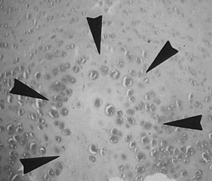
It is obvious from this short summary that histological definitions and descriptions of OC vary, making accurate identification of lesions difficult.24 Even chondrocyte clusters (Figure 54-5), considered by many to be one of the most consistent findings in OC lesions,24,26 are not pathognomonic for the disease,15,24 and, unless used to define the condition, necrosis is not a universal finding.25,27 It is thus not surprising that the relevance to OC of some of the lesions studied has been questioned.28 The matter is complicated further by the limited repertoire of responses that bone and cartilage can mount to injury and developmental abnormalities12 and by the fact that many lesions are not identified until they have reached the chronic stage. The features of such chronic lesions may represent the results of secondary change and attempted repair, rather than primary osteochondrotic change. The results of studies based on such lesions may thus be misleading. This situation has resulted in a wide-ranging, heterogeneous, and somewhat confusing body of literature, in which equine OC has been ascribed to genetic, dietary, endocrine, biomechanical, traumatic, ischemic, and toxic causes.12,18,26,29-33


Relationship among Physeal Dysplasia (Physitis), Subchondral Bone Cysts, and Osteochondrosis


The relationship between OC and physeal dysplasia (physitis or epiphysitis) is poorly defined. Retained cartilage has long been regarded as a possible cause of physeal dysplasia,16,34 and OC lesions of the physis have been described in clinically affected horses (thickened and irregular metaphyseal growth cartilage)5,10 and in foals with experimentally induced dyschondroplasia (retained cartilage cores).26,35 If OC truly is a generalized or multifocal disturbance of endochondral ossification, it would seem logical that metaphyseal growth cartilage has the potential for involvement. However, some have questioned whether articular and physeal lesions have the same cause and have proposed that physitis be regarded as a manifestation of developmental orthopedic disease but not of OC.12 Moreover, the results of a recent study suggest that many enlargements of the distal aspect of the third metacarpal and metatarsal bones, including those with evidence of increased metaphyseal cartilage thickness, represent physiological remodeling rather than a pathological process.36 The role of OC in the pathogenesis of equine physeal dysplasia thus remains unclear. However, it should be noted that a similar pathogenic mechanism (failure of blood supply to growth cartilage) has been proposed for both articular OC and physeal lesions in pigs.1 Physitis is covered in more detail in Chapter 57 and is not discussed further here.


The relationship among OC, subchondral bone cysts, and osseous cystlike lesions is also controversial. In contrast to OCD lesions, which are most commonly found on the nonloaded margins of high-motion joints, bone cysts typically occur in the central, loaded areas of joints.2 Originally interpreted as a manifestation of retained cartilage of the AECC,10,37-39 many cysts are now thought to be traumatic in origin.40,41 This proposed cause was demonstrated experimentally by the successful induction of cysts in the medial femoral condyle (a predilection site) after creation of a linear slit in the articular cartilage followed by normal weight bearing.42 OC may thus be only one of several possible causative mechanisms,12,40,43 and cysts may result from a number of nonspecific articular injuries sustained at a load-bearing site.12


Jun 4, 2016 | Posted by in EQUINE MEDICINE | Comments Off on Pathogenesis of Osteochondrosis

Full access? Get Clinical Tree

Get Clinical Tree app for offline access