Other Soft Tissue Injuries

Chapter 80Other Soft Tissue Injuries


Rupture of the Fibularis (Peroneus) Tertius


History and Clinical Signs


Rupture of the fibularis tertius invariably is caused by trauma resulting in hyperextension of the limb; for example, a horse trying to jump out of a stable and getting one hindlimb caught on the top of the stable door. This usually results in rupture of the tendon in the middle of the crus but occasionally farther distally. Alternatively, rupture may be caused by a laceration on the dorsal aspect of the tarsus, resulting in transection of the tendon. Occasionally, partial tearing of the tendon occurs, usually at the level of the tarsocrural joint, with prominent swelling. Occasionally the reciprocal apparatus is partially but not totally disrupted. Avulsion injuries of the origin of the tendon rarely occur in young foals. imageInjury close to the origin is unusual in mature horses, occurring in two of 25 adult horses with fibularis tertius injury.1


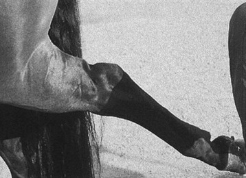
The clinical signs are pathognomonic, because rupture of this tendon allows the hock to extend while the stifle is flexed. When standing at rest, the horse may appear clinically normal, although with acute injury careful palpation may reveal some muscle swelling on the craniolateral aspect of the crus or farther distally. When the horse walks, it should be viewed carefully from behind and from the side. The hock may extend more than usual. The tendons of gastrocnemius and the superficial digital flexor muscles may appear unusually flaccid, and a dimple is seen on the caudal aspect of the crus about one handbreadth proximal to the tuber calcanei. At the trot the horse appears severely lame, with apparent delayed protraction of the limb because of overextension of the hock.


If the limb is picked up and pulled backward, the hock can be extended gradually and “clunks” into complete extension while the stifle remains flexed. A characteristic dimple appears in the contour of the caudal distal aspect of the crus (Figure 80-2). If rupture is only partial, or if lameness is chronic and some repair has taken place, clinical signs may be less severe and the diagnosis less obvious.


Presumably, strain of this tendon can occur, resulting in lameness, but I have no experience of this, and to my knowledge this condition has not been documented.


Diagnosis


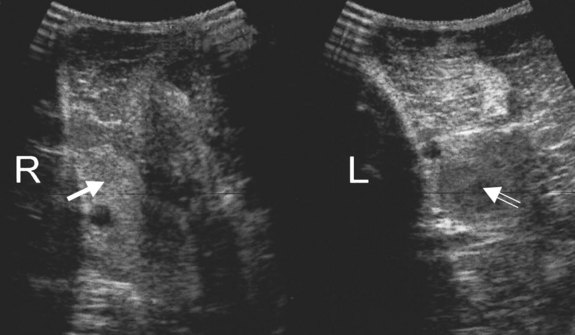
The diagnosis of rupture of the fibularis tertius is based on the pathognomonic clinical signs. The site of rupture can be identified with ultrasonography (see Figure 80-1). The normally echogenic structure is not clearly identifiable and may be replaced by a region that is hypoechoic relative to the surrounding muscles. In horses with chronic injuries the surrounding muscles may become hypertrophied. Usually no associated radiological abnormalities are apparent in adult horses, although avulsion fracture of the origin has been described in foals.

Jun 4, 2016 | Posted by in EQUINE MEDICINE | Comments Off on Other Soft Tissue Injuries

Full access? Get Clinical Tree

Get Clinical Tree app for offline access