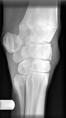
Chapter 17 Traumatic arthritis, osteoarthritis (degenerative joint disease) Osteochondritis dissecans (OCD) Subchondral cyst-like lesions (SCLs) Osteochondroma of the distal radius Osteoarthritis of the small hock joints (‘bone spavin’) Displacement of the superficial digital flexor tendon from the point of the hock/SDFT luxation Rupture of the peroneus tertius • Chip fractures occur at the dorsal margins of the bones forming the antebrachiocarpal and middle carpal joints. Each individual chip fracture affects only one articular surface of the parent bone, although there may be multiple chips affecting different bones, different joints and different limbs in the same horse. The most common sites are the distal margin of the radial carpal bone (Figure 17.1) and the proximal margin of the third carpal bone, i.e. on the dorsomedial margin of the middle carpal joint. The distolateral radius, within the antebrachiocarpal joint is another predilection site. • Slab fractures (see Figure 25.16) extend right through a carpal bone from the proximal to the distal articular surface. They occur most commonly in the frontal plane and primarily affect the dorsomedial aspect (radial facet) of the third carpal bone. Because they involve larger bone fragments and two articular surfaces they are a more serious injury than a chip fracture, and affected horses have a correspondingly more guarded prognosis for return to athletic activity. Slab fractures in the sagittal plane are occasionally seen. • Incomplete fissure fractures of the third carpal bone have been described and may cause lameness with few localizing clinical signs. • Accessory carpal bone fractures are seen most commonly in jumping horses and often there is a history of a fall. The fracture usually occurs in the frontal plane in approximately the middle of the bone. • Comminuted fractures of one or more carpal bones cause severe disruption of the articular surfaces and often result in instability of the carpus. They are usually due to severe trauma such as falls or road traffic accidents. • Lameness localized to the carpus. There may be obvious unilateral lameness or a more insidious bilateral lameness manifested as shortening of the stride and abduction of the distal limb during protraction to reduce the necessity for carpal flexion. • Swelling, especially carpal joint effusion in the early stages, although – as mentioned previously – synovial effusion may not be present in the early stages of subchondral bone disease. Fibrous thickening on the dorsal aspect of the joint occurs with time and obscures the clear contours of the dorsal carpal bone margins. • Pain on joint flexion and sometimes on palpation of the joint margins (especially dorsomedial aspect of Cr and dorsolateral aspect of the distal radius). • Positive response to carpal flexion test. • Restricted range of carpal flexion. Radiology: The four standard weight-bearing projections of the carpus should be supplemented with flexed lateromedial views (to separate the dorsal margins of the radial and intermediate carpal bones) and flexed dorsoproximal–dorsodistal oblique views (to evaluate sclerosis, lysis and fracture of the individual carpal bones) (see Figures 17.1 and 25.55). 1. Traumatic arthritis – see Chapter 15. 2. Osteoarthritis – see Chapter 15. 3. Fractures: most carpal fractures can, in theory, be managed either surgically or conservatively. Surgical intervention is indicated particularly for horses with chip or slab fractures that are intended for athletic use. Surgery aims to restore articular congruency and stability so that the development of secondary OA is minimized. The limitations on this where the fractures are actually a product of existing OA have already been alluded to. • Chip fractures are generally treated surgically by removing the fragments under arthroscopic visualization. Abnormal articular tissues – particularly soft, crumbly subchondral bone – may be debrided at the same time. • Slab fractures are usually treated by lag-screw fixation. Initial debridement and reduction may be accomplished using arthroscopic visualization. • Accessory carpal bone fractures commonly form fibrous rather than bony unions. Techniques for lag-screw repair have been described but are difficult due to the curved shape of the bone and the comminuted nature of many of the fractures. • Comminuted fractures of one or more carpal bones usually end a horse’s athletic career. Salvage for breeding or as a pet may be feasible by combining internal fixation and cast support, with or without combined partial or pancarpal arthrodesis. Comminution of multiple carpal bones is usually an indication for euthanasia. The term ‘angular limb deformity’ refers to lateral (valgus) or medial (varus) deviation of the distal limb from the sagittal plane. Thus ‘carpal valgus’ refers to outward deviation of the distal limb originating at the level of the carpus (Figure 17.2). These deformities are usually seen in young foals and can be congenital or acquired. Figure 17.2 Example of carpal valgus. The distal limb is deviated laterally from the level of the carpus. • Congenital deformities: cause often unknown, but suggestions include malpositioning of the foetus in utero, prematurity/dysmaturity, genetic factors, nutritional influences on the mare, and teratogens. • Acquired: most commonly due to physeal injury or asymmetric loading due to excessive weight-bearing, e.g. as a result of overnutrition or contralateral limb injury, excessive exercise or improper foot trimming. In the case of carpal deformities, the distal radial physis is usually the site affected. • Congenital laxity of the ligamentous support of the carpus and/or hypoplasia (inadequate ossification) of the small carpal bones may result in angular deformity. If left unsupported, the inadequately ossified bones may become crushed and deformed as the foal loads the limbs after birth. • Acquired deformities usually develop as a result of asymmetric growth from the distal radial physis, which may be due to excessive loading of one side of the physis. History: Important facts to establish include: Radiography: It is important to obtain dorsopalmar views centred on the carpus but including as much as possible of the distal radius and proximal metacarpus in order to be able to evaluate the degree of deformity present. 1. Morphological changes: various combinations of the following features may be seen: • Irregularity of the line of the physis; often appears particularly disturbed on the convex side of the deformity. • Metaphyseal and epiphyseal flaring. • Diaphyseal remodelling with asymmetry in cortical thickness. • Hypoplastic, rounded, collapsed, or subluxated carpal bones. 2. The radiographs may also be used to gain an estimate of the degree of angulation present. Traditionally, a piece of clear X-ray film is placed over the dorsopalmar radiograph and lines drawn down the centre of the radius and the centre of the third metacarpal bone. For single-site angulations, the intersection of these lines gives some indication of the level within the carpus at which the deformity is occurring and the degree of angulation (see Figure 25.56). The use of digital radiography and computer software now enables these measurements to be made more easily. Treatment: Some degree of angular limb deformity is not uncommon in neonatal foals and often improves dramatically during the first 2 weeks of life without specific treatment. • Foals should generally be confined to a large box or small yard while the deformity is present, as excessive exercise on an angulated limb tends to exacerbate asymmetric overloading of the physis, carpal bones and other joint structures. • Limb angulation leads to asymmetric wear of the hoof. Foals with carpal valgus tend to wear the medial aspect of the hoof wall excessively. The hoof should be rasped regularly to bring it back into balance. This may be combined with the use of glue-on plastic shoes with medial (in the case of carpal valgus) or lateral (for carpal varus) extensions. • Affected foals should not be allowed to become overweight. This may require restricting the mare’s diet to control milk production, or it may require early weaning. • Neonatal foals with deformity due to ligamentous laxity or carpal bone hypoplasia benefit from external support of the limb with either a well-padded splint or tube cast for 2–4 weeks. Splints should be reset daily and casts changed after 2 weeks to reduce the chance of development of pressure sores. In foals, there is usually little to be gained by external support where there is no evidence of instability, the deformity cannot be reduced manually, and the carpal bones do not appear hypoplastic radiographically. • Foals with persistent deformities due to asymmetric growth from the distal radial physis are candidates for surgical intervention if they fail to respond to the measures outlined above, or if the age of the foal indicates that conservative measures will not have time to be effective. Surgery aims to manipulate growth at the distal radial physis to cause limb straightening. This may involve either: • temporary transphyseal bridging with either staples, screws and wire, or a transphyseal screw, or • There is no need for a second surgery to remove implants. • There is less risk of postoperative infection because there are no implants. Aetiology and pathogenesis: This syndrome is caused by trauma or a space-occupying lesion within the carpal canal leading to compression of the soft tissues within the canal (e.g. accessory carpal bone fracture, osteochondroma of the distal radius, tendinitis of the flexor tendons). It is questionable that this syndrome ever exists as a primary condition and, in the vast majority of cases, is NOT analogous to the human condition, carpal tunnel syndrome, which is associated with nerve compression without a primary lesion. Therefore it is strongly recommended to comprehensively evaluate the horse (using diagnostic imaging and, if necessary, tenoscopy) for an inciting cause. Aetiology and pathogenesis: There is localized abnormal growth of cartilage from an area of metaphyseal periosteum that subsequently undergoes endochondral ossification. It appears to be distinct from the condition of ‘multiple exostoses’, which is thought to be inherited. Clinical signs: Signs are as for carpal canal syndrome plus the bony protruberance on the caudomedial aspect of the distal radius. Osteochondromas are not necessarily a cause of lameness and may thus be an incidental radiological finding. When they do cause lameness, it is often because they impinge on the flexor tendons within the sheath, initially often only at maximal exercise, and hence signs can be intermittent, and therefore diagnosis difficult. Radiology: Radiology shows an irregular exostosis on the caudomedial aspect of the distal radius proximal to the physis; this should not be confused with the normal distal radial physis, which can, in some horses, appear pronounced. Ultrasonography: Inspection of the flexor tendons and the surface of the caudal radius can provide further diagnostically useful information. These usually involve the olecranon process (see Figure 25.8) and commonly the articular surface of the semilunar notch. They compromise the ability of the horse to maintain the elbow in extension during weight-bearing because the fracture interferes with the triceps mechanism of which the olecranon is the insertion. They are subdivided into types according to configuration: Type 1 – physeal fractures occurring in the young horse. Note that if displaced, the epiphysis can move a considerable distance proximally and may even be outside the field of a standard mediolateral radiographic projection of the elbow. Type 2 – Articular fractures involving the semilunar notch. Type 3 – Non-articular fractures involving the metaphyseal region of the olecranon. Type 4 – Comminuted fractures which generally involve the semilunar notch and body of the olecranon. Type 5 – Fractures of the distal olecranon/ulnar shaft that extend proximally to involve the distal semilunar notch. Aetiology and pathogenesis: Ulnar fractures are usually due to a single episode of traumatic overloading, e.g. a kick from another horse or a fall.
Orthopaedics 3. The proximal limbs
17.1 Carpus
Carpal fractures
OA of the carpometacarpal joint
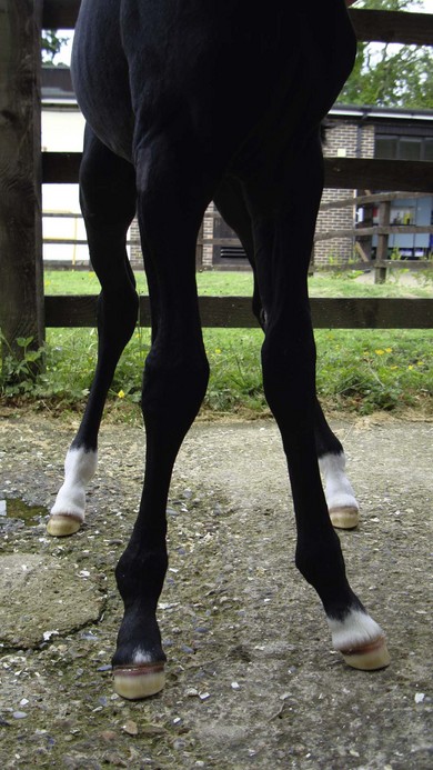
Angular limb deformities
Carpal canal syndrome
Osteochondroma of the distal radius
17.2 Elbow
Ulnar fractures