Chapter 8
Oropharynx and Tonsils
Normal Findings
To correctly identify abnormal criteria, recognition of normal findings is essential. The oropharynx and tonsils are covered by mature squamous epithelial cells (Figure 8-1). These are large, flat, and round to slightly angular. They have abundant pale cytoplasm and small round nuclei that exhibit condensed chromatin. Nucleoli are not visible, and some cells are anuclear. The presence of occasional intermediate squamous cells with slightly larger, less condensed nuclei is normal (Figure 8-2).
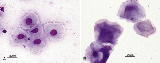
Figure 8-1 Mature squamous cells have cornified cytoplasm that has sharp angular borders. Mature squamous cells may be nucleated or anucleate. A, Mature nucleated squamous cells have abundant light blue-gray cytoplasm that has an angular appearance. B, Mature squamous cell with a pyknotic nucleus (lower left) and anucleate, mature squamous cells from a scraping of oral tissue.
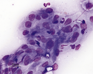
Figure 8-2 Intermediate (less differentiated) squamous cells.
These are also a normal finding from the oropharynx, particularly with samples collected by scraping. Cells are more cohesive and have large, noncondensed nuclei and a more deeply basophilic cytoplasm that does not have angular borders.
The normal squamous cells frequently exhibit a mixed bacterial population adhered to their surfaces (Figure 8-3). These bacteria are also usually present in the background between cells. The normal flora includes aerobic and anaerobic bacterial rods and cocci. Observation of spirochetes is considered normal. Yeast organisms are never considered normal. One bacterium, Simonsiella spp., has a characteristic palisading appearance and is a normal inhabitant of the oropharynx (Figure 8-4). It should never be mistaken for a pathogen. If the bacterial population is dominated by only one type of bacteria, that would be considered abnormal.
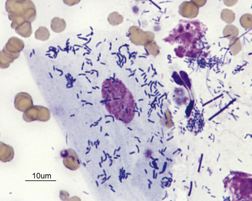
Figure 8-3 Mature squamous cell with adherent bacteria.
Extracellular bacteria are also present. Bacteria adherent to squamous cells and bacteria free in the background of the smear that are not associated with an inflammatory response are usually normal flora.
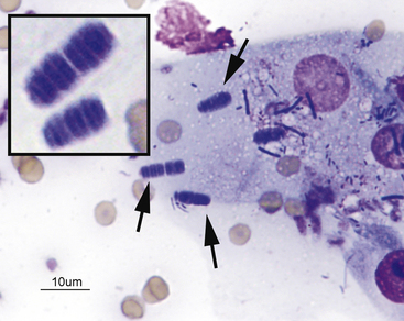
Figure 8-4 Mature squamous cells with Simonsiella spp. bacteria (arrows).
Simonsiella spp. is a normal inhabitant of the oropharynx and must not be mistaken for a pathogen. What appears to be one very large organism is actually numerous slender bacterial rods lined up side to side. Inset, Higher magnification of Simonsiella organisms in which the individual organisms can be seen.
Oropharynx
Nonneoplastic Lesions
Inflammation
Acute (neutrophilic) inflammation is characterized by a predominance of neutrophils. They can be degenerate or nondegenerate. Neutrophils are most frequently degenerate if bacterial endotoxins are present. Macrophages, occasional lymphocytes, plasma cells, fibrocytes and eosinophils can also be present in low numbers.
Infectious agents can be observed; however, their absence from a sample does not rule out the possibility of an infectious etiology. If the inflammatory lesion is caused by a primary bacterial infection or complicated by secondary bacterial infection, a homogeneous population of bacteria is often seen, and many will be phagocytized within neutrophils (Figure 8-5).
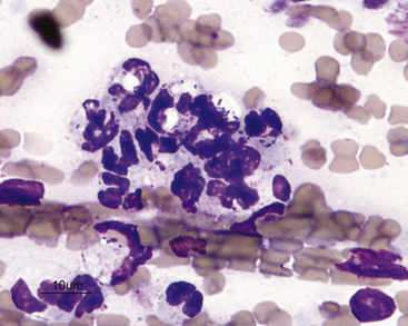
Figure 8-5 Septic, purulent inflammation.
Scraping of a lesion in the oral cavity of a cat shows many neutrophils with phagocytized bacteria. Bacteria that are associated with an inflammatory reaction and are phagocytized by neutrophils likely represent pathogens (primary or secondary).
If the lesion is granulomatous, as from a foreign body or yeast or fungal infection, a more evenly mixed population of neutrophils, macrophages, lymphocytes, and plasma cells is observed, with variable numbers of fibrocytes and fibroblasts that are indicative of physiologic fibroplasia. In some areas of the United States, histoplasmosis in cats can present with oral lesions as the predominant physical examination finding. In these cases, a diagnosis can be made on the basis of identification of organisms from proliferative oral lesions (Figure 8-6).
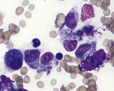
Figure 8-6 Scraping of an oral lesion of a cat with histoplasmosis.
The lesion was pyogranulomatous, yielding a mixture of neutrophils and macrophages. In this image, many macrophages are present and contain phagocytized Histoplasma organisms.
An inflammatory infiltrate, characterized by a predominance of mature lymphocytes and plasma cells with scattered other inflammatory cells, is seen in samples from cats with chronic gingivitis or stomatitis (i.e., lymphocytic–plasmacytic gingivitis or stomatitis). The characteristic inflammatory cells are typically admixed with normal or dysplastic epithelial cells (Figure 8-7).
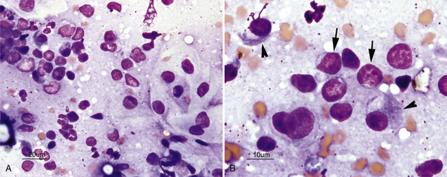
Figure 8-7 Scrapings of a cat with lymphocytic-plasmacytic gingivitis.
A, Low-magnification image shows normal squamous cells (right) and a dense infiltrate of inflammatory cells. B, Higher-magnification image of inflammatory cells shows a predominance of small lymphocytes (arrows) as well as increased numbers of mature plasma cells (arrowheads).
Stay updated, free articles. Join our Telegram channel

Full access? Get Clinical Tree


