CHAPTER 70 Ocular Tumors
Ocular, orbital, and adnexal tumors are rare in cats, affecting 0.34 per cent of all feline patients presented for veterinary consultation.1 These tumors are classified as primary, if they arise in the eye, or secondary, if they involve the eye as a direct extension from a tumor in an adjacent structure (e.g., nasal cavity, brain, or sinuses) or if the tumors are metastatic from another part of the body. In the latter situation, the first clinical evidence of these tumors may be detected in the eye. Ocular tumors can involve the globe, the orbit, or the ocular adnexa (eyelids, conjunctiva, and third eyelid).
PRIMARY TUMORS
EYELID TUMORS
Eyelid tumors are uncommon in cats compared with dogs, occurring in 0.13 per cent of feline patients presented for treatment at veterinary hospitals, and their relative risk increases with age.2 Eyelid tumors in cats are more likely to be malignant than those occurring in dogs; therefore complete surgical resection may require aggressive surgery with or without adjunctive therapy. If the globe is to be preserved, special skin and mucosal flaps are essential to restore the eyelid functionality and maintain the health of the ocular surface. More extensive tumors may require enucleation with skin grafting procedures to obtain adequate margins.3
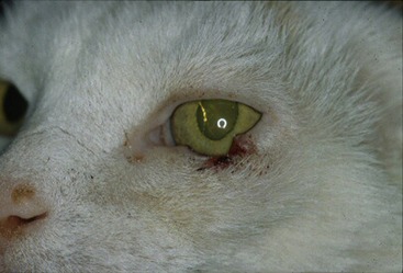
The most common eyelid tumor in cats is squamous cell carcinoma, accounting for up to two-thirds of the tumors in this location.2 This tumor can be found in the eyelids, conjunctiva, nictitating membrane, and orbit, and in advanced cases, it may be difficult to establish the primary site. There is a predilection for the lower eyelid and medial canthus of white cats,1 which suggests that exposure to ultraviolet (UV) radiation and lack of pigmentation may play a role (Figure 70-1). Metastasis usually occurs late in the course of the disease, most often to the regional lymph nodes and lungs4; however, because of the invasiveness of this tumor, local recurrence is common if excised incompletely. As mentioned previously, multiple cutaneous and mucosal flaps may be necessary for restoration of eyelid function.5,6 Other modalities of treatment, in combination with surgery or alone, have been described for squamous cell carcinomas, including intralesional chemotherapy, radiation therapy, photodynamic therapy, and brachytherapy.7–10
Apocrine hydrocystomas or cystadenomas arise from the apocrine sweat glands in the skin of the eyelids or eyelid margin (from the glands of Moll). Results from two recent case reports suggest that these are adenomatous proliferative tumors and not mere retention cysts.11,12 They usually appear in middle-aged to old cats, with a possible predilection for Persian cats because six of the seven cases reported are from this breed.11–13 They usually appear as multiple, well-circumscribed, firm to fluctuant, smooth, pigmented nodules that are 2 to 10 mm in diameter and located in the superior and inferior eyelid, although more typically they arise on the face, neck, and extremities of cats.13 Fine-needle aspirate of these masses usually reveals a dark brown translucent fluid with variably admixed neutrophils or macrophages, and biopsy is needed for definitive diagnosis.11 These proliferations usually are benign, but recurrence and development of new masses may arise at other sites in the eyelids.11,13 Some authors recommend observation without treatment because of the benign nature of the tumor; however, drainage and/or surgical excision are recommended if the lesion is causing discomfort to the ocular structures or resulting in corneal disease.11,13 Chemical ablation with trichloroacetic acid has been suggested as a complement to surgical excision and in the one case report, resulted in no recurrences of the masses 12 months postoperatively.12
Peripheral nerve sheath tumors, also known as schwannomas, neurilemomas, neurogenic sarcomas, neurofibromas, or neurofibrosarcomas, are seen infrequently in the periocular skin of middle-aged to old cats, with a tendency to occur in the superior eyelid.14 These tumors arise as firm nodular masses and, although they usually do not metastasize, local aggressive recurrence after surgical excision is common even when ancillary procedures, such as cryoablation and laser ablation, are used. Radical surgical procedures often are necessary, including enucleation or exenteration of a visual eye.14
Cutaneous hemangiosarcomas involving the eyelid have been described infrequently and in both reported cases involved the lower eyelid.15,16 One case occurred in a white cat, and an association with actinic changes was proposed.16 When compared to its visceral counterparts, cutaneous hemangiosarcomas in cats are associated with a more favorable prognosis17 and have a tendency to recur locally. Metastatic behavior has been reported rarely18; therefore this neoplasm carries a good prognosis when complete surgical resection is achieved.15
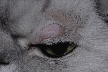
Sebaceous and meibomian gland tumors (adenoma, adenocarcinoma, and epithelioma), so prevalent in dogs, occur rarely in cats.19 Mast cell tumors and melanocytic tumors can occur in the dermal side of the eyelid (Figure 70-2). No specific behavior has been attributed to either of these neoplasms when affecting the eyelids19,20 (see Chapter 67).
CONJUNCTIVAL AND THIRD EYELID TUMORS
Neoplasms of the conjunctiva or third eyelid in the cat are uncommon.19 Conjunctival melanoma has been reported rarely in cats.21,22 Although the number of reported cases of conjunctival melanoma is small, three of the four affected cats were euthanized because of systemic spread of the neoplasm. Local excision was attempted in these patients; however, recurrence of the tumor prompted enucleation. No criteria have been established to predict malignant behavior, and it is unclear if there is any difference if the melanoma arises from the bulbar or palpebral conjunctiva; however, the tumor arose from the bulbar conjunctiva in all three cases in one report.22 These findings suggest that this tumor manifests malignant behavior, necessitating early surgical intervention. A malignant melanoma arising from the nictitating membrane also has been reported,23 with metastases to the brain and lungs. The conjunctiva and orbit were involved extensively, so primary conjunctival or orbital involvement could not be excluded.
Primary hemangioma and hemangiosarcoma of the conjunctiva (bulbar, palpebral, or in the conjunctiva lining the third eyelid) have been reported rarely in cats,24,25 and little is known about their risk factors and prognosis. Distinction between these two tumors is based on histological degree of differentiation and local invasive behavior.20,25 A retrospective study including six hemangiomas and two hemangiosarcomas revealed that six of seven cats in whom gender was reported were neutered males,25 similar to findings for feline cutaneous hemangiomas and hemangiosarcomas.17 Most of the affected patients were middle-aged to old cats. The tumors tended to occur in cats of variably or poorly pigmented hair coats, and the patients lived in states with higher UV radiation levels.25 The most common site of involvement was the leading edge of the nictitating membrane, usually in locations where the conjunctiva was not pigmented. The tumors presented as red-to-brown, smooth-to-multilobulated exophytic nodules. Metastasis appears to be rare, and surgical excision alone was curative in most of the tumors in the series; however, local recurrence may occur when hemangiosarcomas are resected incompletely, necessitating more aggressive procedures such as excision of the entire third eyelid or enucleation. Ancillary therapies, such as laser (neodymium-yttrium-aluminum-garnet [ND:YAG]) and cryotherapy, have been used at the time of excision, but the number of patients in whom these modalities have been used is too low to draw conclusions.24,25
Lymphoma has been reported to occur in the conjunctiva without systemic involvement.26 Another report described a Hodgkin’s-like lymphoma in the conjunctiva of a cat, with various enlarged peripheral lymph nodes; however, histopathological examination was not performed on lymph node tissue to determine if neoplastic tissue was present.27 The cat was reported to be alive 3 years after the diagnosis, after undergoing chemotherapy and radiation therapy.
Adenocarcinoma of the gland of the third eyelid has been reported in one cat.28 The case report described a mass present in the bulbar aspect of the nictitating membrane, causing its protrusion, indentation of the adjacent cornea, and eventually exophthalmos. On histopathological examination, there was a pleomorphic population of anaplastic polygonal cells, with some cells showing intracytoplasmic secretory material and occasional acinar and tubular formations. The cat was euthanized 4 weeks after presentation, with extensive metastatic disease found on necropsy examination. Based on this single case report in the literature, it appears that this tumor has metastatic potential, and radical surgery, such as excision of the nictitating membrane, is recommended at early stages of the disease.28
Mast cell tumors of the third eyelid have been reported in a cat with concurrent eosinophilic conjunctivitis and herpesvirus infection, with no recurrence reported 1 year after local excision.29 Feline cutaneous mast cell tumors usually have a benign course, but it has not been determined if the behavior of conjunctival or third eyelid mast cell tumors in cats differs from their dermal counterparts30 (see Chapter 67).
Squamous cell carcinomas can invade the conjunctiva and third eyelid. The tumors have not been reported as a primary conjunctival neoplasm in cats but rather occur as an extension of eyelid tumors.19
TUMORS OF THE ORBIT
Orbital neoplasms are reported to be rare in cats,31,32 accounting for 4 per cent of feline neoplasms.33 Approximately 90 per cent are malignant,1,31,33 with the average survival time after diagnosis being 1 to 2 months.31,33 The tumors may be primary within the orbit, secondary by extension from adjacent structures (nasal cavity, sinuses), or secondary by metastatic spread,31,32,34 with an estimated percentage of 14, 71, and 14 for each of these categories, respectively.33 These neoplasms tend to occur in older cats, with no breed or gender predilections. The most common clinical sign is exophthalmos, but enophthalmos, protrusion of the third eyelid, epiphora, and strabismus also have been described.31,33 Fine-needle aspirates of these tumors can be useful diagnostically. Advanced imaging techniques (computed tomography [CT] and magnetic resonance imaging [MRI]) have proved to be very useful in the diagnosis of orbital tumors, especially when trying to differentiate these tumors from nonneoplastic diseases.35–37 MRI is less specific for bony structures than CT, but it offers more detailed imaging of the soft tissues of the orbit.38
Epithelial tumors, particularly squamous cell carcinoma, are the orbital tumors reported most commonly in cats, accounting for two-thirds of the tumors in one study.33 In this study, orbital squamous cell carcinoma always occurred secondary by extension from the nasal or oral cavities, conjunctiva, eyelids, or maxillary bone. A variety of other tumors have been reported in the feline orbit, including zygomatic gland tumors,31 adenocarcinoma from the frontal sinuses or nasal cavity,33 lymphoma (associated with systemic involvement or by extension from the nasal cavity),31,33,36 zygomatic osteoma,39 osteosarcoma,31,38,40 fibrosarcoma,31,33,36 melanoma,33 and plasmacytoma41; however, any tissue present in the orbit (including extraocular muscles, adipose tissue, lacrimal gland, and orbital vessels and nerves) can give rise to a primary orbital tumor.
Melanomas have been reported to occur in the orbit by extension from uveal melanomas,33 but only a few cases of primary orbital melanoma without intraocular involvement have been reported.35,42 In both cases, the neoplasms were very advanced at the time of presentation and the possibility of a conjunctival tumor extending into the orbit could not be excluded. Orbital meningiomas, reported commonly in dogs, are extremely rare in cats.31,35
Most orbital neoplasms are malignant. Treatment frequently is unsuccessful because the majority of tumors are secondary to extension from adjacent structures or metastatic from distant sites. Multiple treatment modalities have been described, including surgical excision (usually exenteration of the orbit), radiation therapy, and chemotherapy. Unfortunately, even with the use of multiple treatment modalities, survival time averages 4.3 months.33
Deserving special mention is orbital pseudotumor (retrobulbar pseudotumor, idiopathic sclerosing orbital pseudotumor), a term borrowed from the human literature, which is used to identify any idiopathic mass lesion with accompanying inflammation. It is discussed here because its clinical presentation, behavior, and prognosis are similar to those of a neoplasm.43 This rare condition has been described in the feline orbital area,44,45 occurring primarily in old cats and is characterized by an insidious and progressive course that frequently involves both eyes. The clinical signs usually are related to the orbital disease and include exophthalmos, lagophthalmos, varying degrees of keratitis with possible ulceration and perforation, resistance to manual retropulsion of the globe, and reduced ocular mobility. Histopathologically, the lesion is characterized by an extensive infiltration of the orbit, eyelids, and third eyelid by a variably dense, poorly delineated population of spindle cells (fibroblasts) encircling the eye and dissecting between the orbital adipose and connective tissue, extraocular muscles, and dermal collagen of the eyelids, with variable deposition of collagen and multifocal inflammatory foci. These foci are composed mainly of lymphoplasmacytic infiltrates; however, eosinophilic infiltrates also have been described. Response to treatment (systemic corticosteroids with topical treatment for the secondary corneal disease) in the cases reported is poor, and enucleation is a final outcome in all of the cases reported. Because of the involvement of the contralateral orbit, euthanasia was elected in seven of the eight cases described in the literature.44,45
Stay updated, free articles. Join our Telegram channel

Full access? Get Clinical Tree


