Brian C. Gilger
Ocular Trauma
The horse is subject to a high frequency of ocular and periocular trauma. This is likely because horses’ eyes are large and located at the most lateral aspect of the head (Figure 10-1). This relatively exposed location, coupled with the propensity of horses to move their heads with high speed and force when startled, also likely results in frequent trauma to ocular structures. The clinician should try to ascertain the type of trauma, either blunt or sharp, because these types cause distinct differences in ocular injury. Blunt trauma, such as injury from barn doors, posts, hooves, dirt clods, or whips, is most common, usually causes the most devastating ocular injuries, and carries a poor prognosis for vision. Sharp trauma, by metal nails, aluminum siding, glass shards, or hoof knives, is less common and carries a better prognosis for vision, although the prognosis decreases with increasing depth of penetration of the object into the eye. This chapter is organized according to the inciting cause of ocular injury, either blunt or sharp. In many instances, however, the owner is unaware of the type of injury that occurred. If a thorough history, with discussion of the possible causes of injury, and inspection of the horse’s environment does not lead to ascertaining the underlying cause of the injury, the clinician should assume that the trauma was blunt until proven otherwise. If the cause of the injury is determined, it is important that the owner corrects the problem to prevent repeat injury to this or other horses.
Ocular Consequences of Blunt Trauma
Orbit and Periorbital Structures
Unlike the dog, the horse has a complete bony orbit, composed of the orbital rim anteriorly and orbital walls posteriorly. For this reason orbital bone fractures are sustained more often in horses than in other domestic animals. However, this complete bony orbit provides excellent protection, and other than orbital fractures, the equine eye rarely develops other orbital trauma unless the cornea receives direct impact.
Orbital Fractures
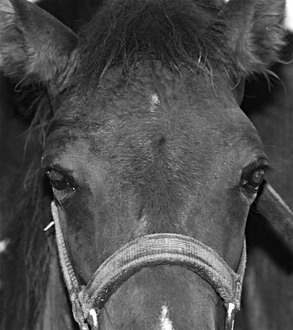
Fractures of the dorsal orbital rim are most common, and the horse is often initially found with a swollen periorbital area, with or without a displaced eye (Figure 10-2). This site is often injured because the rim protrudes externally and laterally and thus is vulnerable to trauma. Other parts of the periorbital rim can also be fractured, depending on the type of trauma. For example, being kicked by another horse may result in ventral orbital rim fractures. Orbital rim fractures may result in displacement, impingement, functional restriction, or laceration of the globe. Fractures from vehicular accidents or from falling over backward and hitting the poll of the head may entail fractures to the basioccipital bone and consequently the basisphenoid bone in the inner orbit. This can result in blindness if the optic nerve is injured or if the globe also directly sustains trauma.
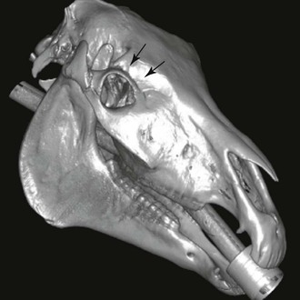
Thorough ophthalmic examination, including digital palpation of the orbital rim both externally and on the conjunctival surface, should be done to diagnose and assess possible orbital rim fractures. For this examination, the horse should be adequately tranquilized and restrained, and after use of topical anesthetic, a lubricated gloved finger should carefully palpate the orbital rim and wall. Placing pressure on the globe or orbital rim should be avoided. Imaging by radiography, and preferably computed tomography, should be performed before surgical intervention is considered. Radiographs of the equine orbit can be taken but are difficult to interpret and rarely adequately demonstrate the extent of the fracture. However, a three-dimensional reconstruction of a computed tomography scan will clearly demonstrate the extent of the fractures and allow proper planning of surgical intervention, if indicated (Figure 10-3). Ocular ultrasound is useful for imaging the ocular posterior segment, including the lens, vitreous, and retina, but does not provide an adequate view of the orbit and orbital fractures. However, ultrasound imaging may help determine whether bone fragments are impinging on the posterior portion of the eye or if there is associated intraocular damage.
Any section of bone that is impinging on the globe or orbital contents should be reduced or removed to prevent damage to the eye. Closed nondisplaced fractures and some closed displaced fractures that are not impinging on orbital structures frequently can be permitted to heal by second intention. For the best cosmetic and visual outcome, closed fracture reduction while the horse is anesthetized is recommended. Zygomatic process fractures may be reduced and closed by manipulation of the bone piece into position by use of a bone hook. More complex fractures of the dorsal orbital rim may be reduced with a malleable plate or bone plate. Fractures should be repaired as soon as possible and at most within 7 days after injury, before development of a callus. Open fractures typically are managed by debridement and cleaning of the wound, reduction of displaced but viable bone fragments, and removal of small, grossly contaminated fragments. Depending on the extent of contamination, some or all the wound is left open for adequate drainage, or drains are placed to facilitate healing.
Periorbital Sinus Disease
Blunt injuries causing periorbital fractures that expose the periorbital sinuses may cause emphysema and epistaxis. Sinus fractures are considered open wounds and should be treated aggressively with antimicrobials and wound care (see Chapter 50). A drain may be placed if there is evidence of infection, such as purulent exudate or cytologic evidence of bacteria or fungus, and creation of drainage should be considered, either externally or into the nasal cavity, if there is inadequate drainage.
Eyelid Lacerations and Contusions
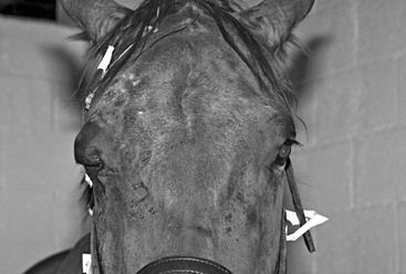
Eyelid trauma and lacerations are very common and usually associated with impact with stall doors, posts, or trailer walls. These blunt injuries result in eyelid contusions and lacerations when the eyelid and associated structures are crushed between the solid injuring structure and the bony orbital rim. Fractures of the orbital rim and periorbital inflammation are also common with these injuries, and complete ocular examination is very important to determine whether other ocular problems such as corneal ulceration, uveitis, hyphema, or retinal detachment are present. Sometimes an eyelid tear (Figure 10-4) is caused by the horse hooking the eyelid on a bucket handle or other metallic structure when the horse startles or panics in a strange situation. This laceration is usually recognized early by the owners and evaluated by the veterinarian when it is still acute.
Surgical correction of nearly all eyelid lacerations is recommended, even if there is substantial damage because loss of the eyelid margin can result in severe, chronic, and vision-threatening keratitis from loss of eyelid protection and tear film components. Because the eyelids have excellent blood supply, most lacerations can be repaired to achieve relatively well-functioning and cosmetic eyelids. Hanging eyelid pedicles should not be removed, and wound debridement should be minimal during surgery. A two-layer closure with an initial deep subconjunctival layer of a continuous absorbable suture, such as 6-0 polyglactin 910, will ensure that the conjunctival aspect of the eyelid does not gape during healing and induce scar formation. Inadequate closure of the conjunctiva resulting in scar formation, and poorly placed sutures (e.g., full-thickness conjunctival sutures) resulting in corneal trauma, are the most common causes of dehiscence and complications, respectively. The eyelid margin is reapposed meticulously by first suturing the margin, preferably with a figure-of-eight suture pattern of nonabsorbable 4-0 to 6-0 suture. Additional simple interrupted skin sutures are placed as needed to close the remainder of the eyelid defect. Skin sutures may be removed in 10 to 14 days.
Corneal Injuries
Corneal Ulceration
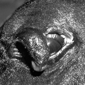
A corneal ulcer is a break in the corneal epithelium that exposes the corneal stroma. In the horse, this is usually caused by trauma. Usually, owners recognize this lesion by its acute onset and frequently by the accompanying severe lacrimation, blepharospasm, photophobia, conjunctival hyperemia, and ocular cloudiness. Corneal ulceration can be a result of blunt or sharp trauma. With blunt trauma, the corneal lesion is usually superficial; however, associated intraocular damage such as uveitis, hyphema, vitreal hemorrhage, and retinal detachment is common. A thorough and complete ophthalmic examination is required, including ocular ultrasound if the ocular posterior segment cannot be viewed because of corneal edema or miosis. Furthermore, the conjunctival cul-de-sacs and area beneath the third eyelid should be examined thoroughly for foreign bodies or environmental debris. Removal of foreign material is essential for healing of the corneal ulcer. Because the horse’s eye is very painful, tranquilization, local anesthesia of the eyelids, and use of topical anesthetic (1.0% proparacaine HCl) and a vasoconstrictor (2.5% phenylephrine HCl) may be necessary to permit thorough examination. In most horses, treatment with a topical broad-spectrum antimicrobial (e.g., neomycin, bacitracin, and polymyxin B solution or ointment) every 6 hours, topical atropine HCl ophthalmic 1% every 12 to 24 hours, and systemic flunixin meglumine (1.1 mg/kg, IV or PO) will allow rapid healing of the corneal ulcer. The horse should be reevaluated every 2 to 3 days until epithelium covers the ulcer. For more information on the diagnosis and treatment of corneal ulceration, see Chapter 143.
Corneal Edema
Acute onset of corneal edema may result from blunt trauma to the eye. There is usually associated intraocular inflammation; therefore thorough ophthalmic examination, including measurement of intraocular pressure, is warranted. There is no specific treatment for corneal edema, except to treat intraocular inflammation.
Stay updated, free articles. Join our Telegram channel

Full access? Get Clinical Tree


