Chapter 64 Ocular Disease and Suspected Causes in Captive Pinnipeds
Ocular disease is one of the most common medical problems observed in captive pinnipeds, with a disproportionally higher prevalence when compared with similar eye problems in their wild counterparts or other captive mammalian species. Possible causes, both studied and anecdotal, are one or a combination of factors, including trauma, infection, periodic or persistent exposure to excessive chemical, oxidant, or noxious byproduct levels in the water, osmolality of the water, viral or bacterial pathogens, excessive ultraviolet (UV) light exposure, nutritional imbalances, and genetic predisposition.5–7 When single or multiple pinnipeds develop recurrent eye problems, a comprehensive review of possible causative factors is advised. In addition to an ophthalmic examination, a thorough evaluation of oxidant levels in the pool, coliform counts, environmental conditions, including pool color and availability of shade, salinity, nutritional supplements, exposure to pathogens, and prevalence of trauma is warranted.
Ocular Disease
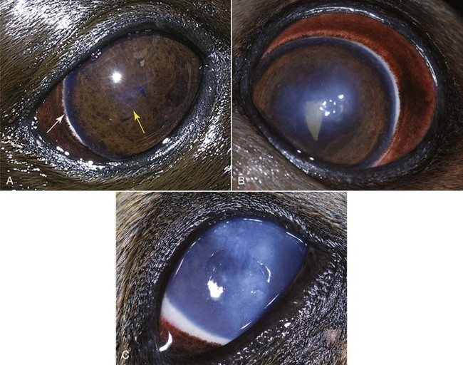
Corneal disease is a painful and frequently recurrent problem in captive pinnipeds. Many have experienced mild to moderate ulcerative corneal disease and may become visually impaired by the time they reach their teenage years. Common corneal problems seen in pinnipeds are edema, opacities, and chronic keratitis. In one 6-year study conducted on 113 captive otariids in North America and the Bahamas, keratitis was identified in one or both eyes of more than 45% of the animals evaluated.1 Flare-ups occurred in the most affected animals two to four times/year, primarily during seasons when the sunlight exposure increased or, in tropical climates, became more intense. In winter, flare-ups were seen when there were bright sunny days, with snow continuously on the ground. In two study facilities in which animals lived primarily indoors, the resident pinnipeds had less severe corneal disease for their age and fewer annual flare-ups of keratitis. This study defined three stages of keratitis in otariids and identified this common syndrome as otariid keratitis (Fig. 64-1).1 Repeated insults to the cornea could lead to secondary bacterial or fungal infections. When trauma and primary infectious processes are ruled out, frequent oxidant or noxious byproduct spikes in the water and excessive exposure to UV light are two of the more likely causes. An ongoing large-scale epidemiologic survey involving 20 facilities worldwide will help determine whether sun exposure is truly a risk factor for this disease.1
Another study has described cataracts or lens luxations in pinnipeds older than 5 years, which became increasingly more common in older animals. In a study group of 111 pinnipeds, the prevalence of cataracts, with or without lens luxation, was 21% in those aged between 6 and 10 years 58% in those between 11 and 15 years, 66% in those in the 16- to 20-year-old age group, 87% in animals between 21 and 25 years old; all 9 study animals older than 26 years had cataracts, and 5 of them also had lens luxations.2 The risk factors for developing cataracts, lens luxations, or both that were identified in this study included increasing age, history of ocular disease, history of fighting, and lack of shade. Animals in this study without access to shade were 10 times more likely to develop cataracts or lens luxations.2
Environmental Issues
Pool Color and Ultraviolet Light
Historically, many pinniped exhibit pools were painted a light blue color, and many exhibit pool and holding area walls are a light color or natural concrete. Most animals included in the cataract study were housed in these conditions for most of their lives. Efforts to create more naturalistic exhibits and paint pool surfaces tan or other less reflective colors have been more recent. Although pool or exhibit color was not directly identified in the cataract study as a risk factor for developing lens luxations or cataracts, the lack of shade was significant.2 This suggests that chronic excessive exposure to UV light is likely a factor in the early development of cataracts and/or lens luxations in pinnipeds. It might be argued that pinnipeds have evolved in a bright ocean environment, but wild pinnipeds are not exposed to the same amount of UVA and UVB light as many of their captive counterparts.
Comparing captive pinniped housing and behavior, which may contribute to the amount of UV light exposure that they experience over time, with that of wild pinnipeds may be revealing. Captive animals may be held in bright reflective pools, many of which are painted a light blue color. Although these bright pools may exhibit the animals nicely, they reflect a considerable amount of UV light back into the animals’ eyes. Wild pinnipeds swim and feed in a nonreflective environment, looking downward into deeper darker water as they hunt for food. The ocean floor has a relatively nonreflective surface. Captive pinnipeds are more inclined to look up frequently toward the sky than their wild counterparts. Captive animals are usually fed by trainers holding fish for the animal to eat or by keepers who broadcast-feed the animals by throwing quantities of fish into the pool. Some facilities allow the public to feed the animals by having them throw or drop fish into the exhibit pool. In all these cases, the captive animals orient their eyes skyward to locate or receive their fish reward. In some cases, keepers or trainers feed the animals from one location in the exhibit throughout the day. Care should be taken to ensure that the animal never has to look directly into the sun when being fed. Wild animals may haul out on bright reflective beaches, but they typically rest or sleep with their eyes closed. They may occasionally look about their surroundings or at one another but rarely gaze skyward. Captive animals are frequently surrounded by natural concrete or light-colored walls in their exhibit or holding area. The combination of the light-colored reflective pools and the fish being fed from above (“sky fish”) causes them to receive considerably more UV light exposure than their wild counterparts. Studies in humans and other animals have suggested that progressive eye damage may be the result of excessive UV light exposure.3,4,9 A recent study has found that pinnipeds with lack of shade are 10 times more likely to develop cataracts and/or lens luxations.2 This may explain why I have observed that captive pinnipeds held in U.S. facilities at latitudes below 33 degrees N with a preponderance of bright sunnydays, housed in light blue salt water pools, with no access to shade, appear to have a greater prevalence of clinically apparent eye disease than animals held indoors or in northern latitudes, especially considering that some of these sunny facilities have few, if any, water quality issues.
Stay updated, free articles. Join our Telegram channel

Full access? Get Clinical Tree


