Chapter 5 Observation
Symmetry and Posture
Assessments of symmetry and posture are important aspects of a lameness examination. Comparison between the normal and abnormal sides facilitates identification of abnormalities, unless the condition is bilateral so that no recognizable differences exist between the left and right limbs. The horse should be standing squarely on a flat surface in a quiet, insect-free environment. Horses with severe lameness often are reluctant to stand correctly, but information gained about symmetry and posture of severely lame horses is valuable. The veterinarian should look carefully at size, shape, contour, heights, and widths and compare with the opposite side.
Forelimb Symmetry
Muscle Atrophy
The symmetry of skeletal muscle in the forearm, pectoral, and cervical areas should be assessed. Muscle atrophy that occurs in horses with chronic lameness conditions is called disuse atrophy and in those with neurological disease is called neurogenic atrophy. Horses with muscle atrophy and lower motor neuron disease (see Chapter 11) may be lame, sometimes as a result of muscle pain or nerve root pain, complicating differentiation between these causes of muscle atrophy. In most but not all horses with neurogenic atrophy, other clinical signs suggestive of neurological disease may be present. Horses with disuse atrophy resulting from chronic lameness usually have generalized atrophy of the ipsilateral forelimb. Muscle loss usually is not pronounced but involves the forearm (extensors are most commonly affected), triceps, and shoulder muscles. Shoulder muscle atrophy involving the infraspinatus and supraspinatus muscles generally is not pronounced, and lateral subluxation of the shoulder joint during weight bearing is not present (see Chapter 40).
Horses with neurogenic atrophy may have profound atrophy of one or more muscles in the forearm, pectoral, or cervical regions. Atrophy often is much more pronounced than expected based on the degree of lameness, prompting suspicion of neurological disease. Pronounced, unilateral pectoral or triceps atrophy with mild atrophy of the forearm muscles suggests neurological disease. Severe atrophy localized to the infraspinatus or supraspinatus muscles without subluxation of the shoulder joint usually results from injury of the suprascapular nerve caused by external trauma. Atrophy and subluxation of the shoulder joint is associated with injury of the brachial plexus or nerve roots. Other muscles may also show atrophy.
Localized muscle atrophy or fibrosis occurs in horses with previous injury and subsequent scar tissue formation within muscle bellies. This condition is more common in the hindlimb but occasionally occurs in the forelimb.
Swelling
Swelling, a common sign of inflammation, often causes asymmetry. The presence of swelling and heat should alert the lameness diagnostician to the possibility of an infectious process and may lead to additional examinations such as assessment of body temperature and the acquisition of laboratory data. Swelling within a joint capsule caused by excess joint fluid, effusion, is a general reaction of the joint to several traumatic or degenerative processes. Edema, cellulitis (lymphangitis), bleeding, and fibrosis can cause soft tissue swelling. Underlying bony enlargement can mimic soft tissue swelling, particularly in older horses with advanced osteoarthritis. Some swellings are clinically innocuous, such as mild distention of a digital flexor tendon sheath (so-called “windgalls” or “wind puffs”), but a recent change in size, local heat, or marked left-right asymmetry should alert the clinician to a possible problem. Occasionally a well-circumscribed tense spherical swelling is seen palmar to the neurovascular bundle on the side of the fetlock. These synovium-filled masses are usually incidental findings of no clinical significance. Edema usually signals acute inflammation, and pits (a distinct impression is visible) when compressed by digital palpation (pitting edema). Horses develop edema around and often distal to the site of inflammation. In some horses, especially racehorses left unbandaged when accustomed to being bandaged, benign mild-to-moderate edema of the distal extremities develops. This process is called “stocking-up” and should not be misinterpreted as a pathological process. In these horses the edematous area is not painful and usually does not pit, and the horse is not lame. Edema in these horses can complicate lameness examination because it is sometimes difficult to palpate underlying structures and to perform diagnostic analgesia.
Cellulitis describes infection within the tissue planes of the distal extremities (see Chapter 14) and is sometimes called lymphangitis. Lymphangitis, by definition, is inflammation of the lymphatic circulation of the limb, but the conditions are similar and the terms are used interchangeably. Swelling is firm, warm, and painful, and lameness is often pronounced. “Stovepipe” swelling describes this condition (“the horse is all stoved-up”). Horses generally show systemic signs such as fever and elevated white blood cell count. Cellulitis usually results from small puncture wounds that may be difficult to discover or occurs after articular, periarticular, or subcutaneous injections. Infection develops in subcutaneous tissues or deeper in the dense fascial planes and can be difficult to eradicate.
Fibrosis or scar tissue formation as the result of previous cellulitis or trauma causes asymmetry of the distal extremities but may not be the source of the current lameness. The veterinarian should avoid overinterpreting areas of scar tissue formation unless evidence of recrudescent inflammation exists. Horses may have scars caused by previous application of counterirritants or from healed wounds, leaving large, painless, and thus benign blemishes. Scars from previous surgical procedures, sometimes recognized by small areas of white hair accumulation, should be noted but may have no bearing on the current problem. Previous scars may have more relevance during prepurchase examinations.
Bony swelling is a common cause of asymmetry. Proliferative change results in periosteal or periarticular new bone formation and accompanies myriad problems in the distal extremities. Bony changes may be active, causing the current lameness problem, or old and inactive, causing few or no clinical signs. For example, old inactive bony swelling of the shin or osselets (bony and fibrous swelling of the fetlock joint) may be prominent in ex-racehorses but may have little to no relevance to current lameness.
Angular Deformity
Angular limb deformities in young horses are common, are sometimes associated with lameness or other developmental orthopedic disease, and are discussed elsewhere (see Chapter 58). Abnormalities of conformation should be noted but may have little relevance to the current lameness problem (see Chapter 4). Horses younger than 2 years of age with severe forelimb lameness of several months’ duration may develop contralateral varus deformity originating from the carpus or elbow joints.
Foot Size
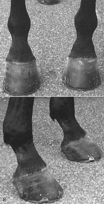
Ideally both front feet should be identical in size and shape, or nearly so, and any asymmetry should be noted. Horses with chronic lameness may have disparity in foot size, usually with the smaller foot being ipsilateral to lameness. The small foot often is contracted and more upright (Figure 5-1). Chronic reduction in weight bearing results in foot size disparity in some, but not all, horses. Mild disparity in foot size is a normal finding in some horses. Mild clubfoot conformation, acquired from previous flexural deformity, may be present incidentally in adult horses. Previous lameness may have caused contraction of the foot but has since resolved, resulting in disparity in foot size and shape but no residual lameness. In these horses it would be a mistake to assume current lameness is originating from the foot without confirmation using diagnostic analgesia. Clubfoot conformation appears to be better tolerated in Thoroughbred (TB) than in Standardbred (STB) racehorses.
Fetlock Height
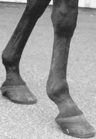
Fetlock position should be assessed in the standing horse and during movement. In a standing horse, fetlock height should be symmetrical, assuming the horse is loading the limbs equally. Horses with severe lameness commonly “point” or hold the limb in front of the opposite forelimb, thus taking weight off the limb. This standing posture obviously causes disparity in fetlock height but should be carefully interpreted. Loss of support of a fetlock in the standing horse causes the affected fetlock to drop and occurs most commonly with acute, traumatic disruption of the suspensory apparatus in racehorses but also appears with chronic, active desmitis (Figure 5-2). Severe superficial digital flexor tendonitis or lacerations resulting in fiber damage of the deep or superficial digital flexor tendons can cause similar clinical signs.

Fig. 5-2 Standardbred racehorse with severe suspensory desmitis and a “dropped fetlock.” The level of the right front fetlock joint is lower than that of the left front, caused by chronic, severe desmitis. Similar clinical signs and severe lameness appear in horses with acute traumatic disruption of the suspensory apparatus.
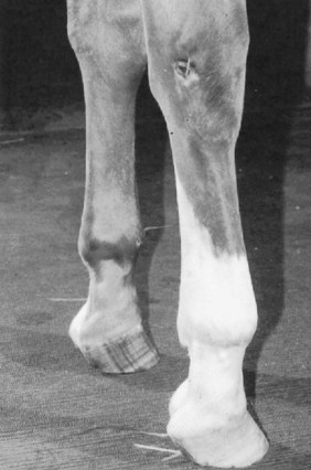
In horses with mild flexural deformity of the metacarpophalangeal joint, dynamic knuckling (buckling forward, flexion) of the fetlock joint may occur in the standing position (Figure 5-3). Joint position usually returns to normal during movement. In horses with severe flexural deformity, normal fetlock position is never achieved. Knuckling of the fetlock also may result from desmitis of the accessory ligament of the deep digital flexor tendon.

Fig. 5-3 Knuckling forward of the right front fetlock joint occurs in a standing position in this horse with mild flexural deformity of the metacarpophalangeal joint. This dynamic instability abates somewhat when the horse moves, but the left front fetlock also is straight, indicating the presence of bilateral flexural deformity.
Scapular Height
Disparity in scapular height is a rare clinical sign in a lame horse. The veterinarian must stand behind and above the horse to observe scapular height. The horse’s mane may obscure observation from a distance, requiring closer examination by palpation. Traumatic or neurological conditions affect scapular height, causing either injury or dysfunction of the serratus ventralis muscle, respectively. With both conditions the dorsal aspect of the scapula is higher on the affected side. The veterinarian may place pieces of white tape or other suitable markers on both sides of the horse and stand back to compare height or may use two assistants to point to the locations. Horses may have disparity in scapular height unrelated to the current lameness condition if there has been resolution of the original injury or neurological problem. Care must be taken to differentiate between genuine differences in the height of each scapula and asymmetrical musculature, which occurs much more commonly and may be an incidental observation. Horses with disparity in scapular height or asymmetrical muscle development may have problems with saddle fit.
Hindlimb Symmetry
Muscle Atrophy
Asymmetry of bone and muscle mass in the hindlimbs and pelvis is a common clinical sign but must be differentiated carefully. The horse should stand squarely on a flat, even surface. The clinician must determine whether asymmetry exists, and if so, if the problem involves muscle, bone, or a combination of the tissues. Muscle atrophy is most common and, if unilateral muscle atrophy exists, easily can be confused with bony asymmetry caused by pelvic fractures or asymmetry of the tubera sacrale.
Disuse and neurogenic muscle atrophy occur in the hindlimb. Horses with chronic hindlimb lameness develop ipsilateral gluteal muscle atrophy, but asymmetry may be subtle. Mild muscle atrophy usually first appears just lateral to a tuber sacrale. The veterinarian should differentiate muscle atrophy from disparity in height of the tubera sacrale (Figure 5-4

Stay updated, free articles. Join our Telegram channel

Full access? Get Clinical Tree

